SNT Gatchaman

Senior Member (Voting Rights)
Staff member
Heightened innate immunity may trigger chronic inflammation, fatigue and post-exertional malaise in ME/CFS
Xiaoyu Che; Amit Ranjan; Cheng Guo; Keming Zhang; Rochelle Goldsmith; Susan Levine; Kegan J. Moneghetti; Yali Zhai; Liner Ge; NISCHAY MISHRA; Mady Hornig; Lucinda Bateman; Nancy G. Klimas; Jose Gilberto Montoya; Daniel L. Peterson; Sabra L. Klein; Oliver Fiehn; Anthony L. Komaroff; W. Ian Lipkin

Myalgic encephalomyelitis/chronic fatigue syndrome (ME/CFS) is characterized by unexplained fatigue, post-exertional malaise (PEM), and cognitive dysfunction. ME/CFS patients often report a prodrome consistent with infection.

We present a multi-omics analysis based on plasma metabolomic and proteomic profiling, and immune responses to microbial stimulation, before and after exercise. We report evidence of an exaggerated innate immune response after exposures to microbial antigens; impaired energy production involving the citric acid cycle, beta-oxidation of fatty acids, and urea cycle energy production from amino acids; systemic inflammation linked with lipid abnormalities; disrupted extracellular matrix homeostasis with release of endogenous ligands that promote inflammation; reduced cell-cell adhesion and associated gut dysbiosis; complement activation; redox imbalance reflected by disturbances in copper-dependent antioxidant pathways and dysregulation of the tryptophan-serotonin-kynurenine pathways.

Many of these underlying abnormalities worsened following exercise in ME/CFS patients, but not in healthy subjects; many abnormalities reinforced each other and several were correlated with the intensity of symptoms. Our findings may inform targeted therapeutic interventions for ME/CFS and PEM.

Web | PDF | Preprint: MedRxiv | Open Access
 
That's a vague abstract - makes me wonder if they found anything really important.
Good to see something from Columbia. 56 ME/CFS participants; 52 healthy controls

Results said:
Study subjects were 56 ME/CFS cases and 52 HC recruited in New York and California, with 47 case-control pairs matched for sex, age (±5 years), race/ethnicity, geography, date-of-exercise and blood sampling (±12 weeks), diagnosed using the CDC/Fukuda and Canadian consensus
criteria.1,6

Heightened cytokine responses to SEB and HKCA in ME/CFS

Results said:
We used the TruCulture ex-vivo system7 to ask whether expression of inflammatory mediators, before and after exercise, differed between ME/CFS and HC following exposure to superantigens and mimics of viral, bacterial and yeast infection: Staphylococcus aureus enterotoxin type B (SEB), heat-killed Candida albicans (HKCA), lipopolysaccharide (LPS) and polyinosinic:polycytidylic acid (poly I:C).
I'm interested to know what people think about this TruCulture system. Presumably things would be quite different actually in the body, where signals might be coming from muscles and other parts of the body as a result of infection, and interactions might happening with other cells. But, I guess it's still worth a look. It just means that if there isn't something found ex-vivo, that doesn't rule out something happening in the body.


SEB - Staphylococcus aureus enterotoxin type B SEB
Results said:
In ME/CFS, levels of CXCL5, GM-CSF, IL-1β, IL-2, IL-6, IL-8, IL-23, and TNF-α were elevated before exercise in blood exposed to SEB (Fig. 1A, Table S8E). Similar elevations were seen with IFN-γ, IL-13, and IL-17 (padj<0.10). SEB drives T-cell expansion by crosslinking T-cell receptors (TCRs) with major histocompatibility complex class II molecules (MHCIIs). 8 ME/CFS cytokine profiles indicate elevations in superantigen-induced T-cell-mediated immune responses.
Screenshot 2025-07-25 at 3.14.05 pm.png

The two orange bars are ME/CFS before and after exercise; the blue and grey bars are healthy controls before and after exercise. SEB is . At first look, there are major differences. But, there are some outliers in the healthy controls, perhaps some bias in the treatment of outliers? And the means aren't actually all that different, and the p values aren't that strong. Perhaps there is a subset of ME/CFS where expression of the cytokines increases more in response to the SEB? There doesn't seem to be a lot going on with respect to changes as a result of exercise in either cohort except in healthy controls with IL-23.

They looked at differences between ME/CFS and HC in various subsets (females, males, females <45 years, females> 45 years). They note that cytokine responses were greater in females. I'm not seeing much of interest there. Given males and females, young and old, get ME/CFS symptoms, I don't think much should be made of different results in these subsets.


HKCA - heat-killed Candida albicans
Results said:
In response to HKCA, ME/CFS subjects had elevated levels of IFN-γ and TNF-α before exercise compared to controls. Baseline levels of IL-1β were also elevated but did not reach significance (padj=0.073). The pre-exercise cytokine elevations in response to HKCA were similar in females and males with ME/CFS (Fig. 1D).
Figure 1C

Screenshot 2025-07-25 at 4.09.14 pm.png

So, mean baseline levels of IFN-y and TNF-a do look different between ME/CFS and HC. But, there's a lot of overlap and outliers. And values normalise 24 hours after exercise.


Discussion said:
In the TruCulture ex vivo system, immune stimulation with SEB and HKCA induced higher levels of pro-inflammatory cytokines in ME/CFS than in HC with sex- and age-specific effects. Although some findings did not reach statistical significance (padj<0.10), the pattern of the data was consistent across pro-inflammatorycytokines (Fig. 1A, 1B). We posit that these differences were influenced by E2, a master regulator of inflammation inhibiting the release of pro-inflammatory cytokines, including IL-1β, IL-6, and IL-23. E2 also promotes expansion of Treg cells, and inhibits differentiation of Th17 cells and production of Th17 cytokines9
It is not clear to me how 'E2 the master regulator of inflammation' is causing the differences found.

They say they found higher levels of pro-inflammatory cytokines in ME/CFS and then say that E2 inhibits the release of pro-inflammatory cytokines. It's not clear if they think E2 is doing what it should or not. If it is inhibiting the release of things like IL-6, or even not inhibiting the release of them, that's hard to square with quite similar mean levels of IL-6 in the ME/CFS and HC cohorts for both the SEB and HKCA experiments.

For IL-23, mean levels are a bit higher in ME/CFS when cells are stimulated by SEB, but Il-23 doesn't even make it onto the chart for the HK Candida stimulated samples.

So, I'm not sure yet if they are suggesting that E2 the master regulator of inflammation is causing an expansion of Tregs in ME/CFS, or if the problem is, it is not.


I'm finding this all a bit hard going and opaque. And, there's a lot in this paper. This is just the first section.
 
Onwards...

Sensitivity of cytokine responses to LPS and poly I:C
Results said:
TruCulture preparations exposed to LPS and poly I:C revealed higher secretion of pro-inflammatory cytokines compared to no stimulation, in both ME/CFS and HC (Table S1C). However, we did not observe case-control differences (Fig. S1, Table S8B & S8D).
So, no differences between healthy controls and ME/CFS when PBMC were stimulated with LPS or poly I:C (proxies for bacterial and viral infections respectively).

The authors thought this lack of difference might be due to the limitations of their TruCulture system (specifically "saturation of their cognate Toll-like receptors"). They had a problem with not having enough samples, but did a small study (ME/CFS=8; HC=7) with different concentrations of LPS and poly I:C. They say about this
We observed dose- and time-dependent IL-6 responses to both LPS and poly I:C stimulation at 12, 24 and 48 hours of incubation
Which is again not very specific or helpful.

Looking at charts 1E and 1F, it does look as though the ME/CFS cells from pre-exercise samples might be producing quite a lot more IL-6 than the HC cells when incubated for longer at the higher concentrations. e.g. more IL-6 with 100 ng/ml of LPS incubated for 24 hours than with 0.01 ng/ml of LPS. Note the range bars - very large for the LPS ME/CFS samples, and for one rather anomalous HC experiment, and note the outliers that don't get included.

Screenshot 2025-07-25 at 4.47.25 pm.png

Discussion said:
Accordingly, we tested IL-6 responses in PBMCs exposed to various doses of LPS or poly I:C for 12, 24 or 48 hours. These experiments revealed differences in innate immune responses that were most evident at lower stimulant concentrations and at 24 hours of incubation.
I think that statement in the Discussion is wrong if Chart 1E is correct. From the chart, the biggest difference is with the highest concentration of LPS incubated for 48 hours. Perhaps I am misunderstanding something?
 
I think studying blood cells in culture like this and looking for increased responsiveness that might indicate "priming" is a valid way to test theories about what that priming might then cause. But they wreck the whole story by a title that suggests that what it would cause is chronic inflammation when that is the one thing that isn't there in ME/CFS.

It would be interesting to know what cells were primed and what they might then respond with in relation to some theory about them arriving somewhere and doing something - but there has to be a meaningful theory of what they are then expected to do where.

I am finding it hard to interpret the data but it looks as if exercise had no effect on any of this? If so why not state that clearly as an important finding?
 
How important is it likely to be that they didn't have sedentary controls?

Also, I was surprised to see them say, 'Cells from muscle and other metabolically active organs are not available from ME/CFS patients'. That's surprising. Is it true in the UK? Do we need a cell/tissue biobank?
 
Wrote a short summary, highlighting some findings:


1) An impressive dataset on ME/CFS was just published by the research team of Ian Lipkin. They tested multiple proteins and metabolites in 56 ME/CFS patients and 51 controls before and after exercise and cytokines in response to mimics of viral, bacterial, and yeast infection.

2) Let's start with the cytokine response. The researchers measured this after exposure to antigens that mimic:
- a fungal infection (HKCA)
- a bacterial infection (LPS)
- a viral infection (poly I:C)
- superantigens (SEB) which triggers a nonspecific T-cell response

3) No group difference were found for the bacterial and viral exposure, some differences were seen after HKCA (fungal) while the biggest differences were found for the superantigen SEB.The cytokine response to SEB was much higher in patients compared to controls.
1753442371405.png

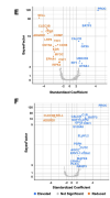
4) Overall, the exercise test did not seem to have a dramatic effect; there were often as many difference between patients and controls before (Figure 2.E) versus after exercise (Figure 2.F). But the differences after exercise were different more often increases compared to HC.
1753442382113.png

5) One interesting result was complement activation after exercise which has been reported in ME/CFS before. Here, levels of C1R and CFHR4 were elevated.

The authors also note abnormalities in calcium signaling with increased levels of CALCR, RAMP3, and dS100A8 after exercise.

6) There were too many results to highlight them all. Picking out some findings, the authors found lower plasma levels of SELL, CDH5, ADGRD1, and KDR which suggest impaired immune surveillance and dysregulated immune activation.

7) ME/CFS patients also had lower levels of carnitines, particularly acylcarnitine (CAR) after exercise. The authors found reduced baseline CAR in earlier studies as well.

Kynurenine/Tryptophan (TRP) ratios were lower in ME/CFS before exercise but increased after exercise.

8) Another interesting finding was reduced levels of proteins produced by neuronal cells, such as EPHA4 and CNTN4. These modulate signal transduction at synapses, which fits with a recent genetics study that also pointed to synapse communication.
https://www.medrxiv.org/con.../10.1101/2025.04.15.25325899v2

9) Some caveats about the study: the sample size was relatively small for the many complex analyses done. And although the authors adjusted for important covariates, they admit that they cannot rule out diet, medications, and lifestyle as potential confounding factors.
 
8) Another interesting finding was reduced levels of proteins produced by neuronal cells, such as EPHA4 and CNTN4. These modulate signal transduction at synapses, which fits with a recent genetics study that also pointed to synapse communication.
Here's what this paper says about this:
Plasma proteomic findings also revealed abnormalities in proteins predominantly produced by 290 neuronal cells. Before exercise, levels of Ephrin receptor A4 (EPHA4) and contactin 4 (CNTN4) 291 were lower in ME/CFS (Fig. 2E). EPHA4 is highly expressed in brain regions with high synaptic plasticity and is implicated in learning and memory.54 292 CNTN4 is expressed in both peripheral and central nervous system and is involved in modulating signal transduction at synapses.55 293294 After exercise, levels of ELAV-like RNA binding protein 2 (ELAVL2) were elevated (Fig. 2F). 295 ELAVL2 regulates the post-transcriptional expression of key genes involved in neuronal development and function.56 296 In response to exercise, levels of Neurexin1 (NRXN1) were 297 decreased in ME/CFS (Table S6A), indicating lower synaptic adhesion and neuronal 298 transmission that may contribute to PEM-associated cognitive impairment.
 
5) One interesting result was complement activation after exercise which has been reported in ME/CFS before. Here, levels of C1R and CFHR4 were elevated.
Some previous studies focused on c4a but results were not entirely consistent:
Complement activation in a model of chronic fatigue syndrome - PubMed
Unravelling the nature of postexertional malaise in myalgic encephalomyelitis/chronic fatigue syndrome: the role of elastase, complement C4a and interleukin-1beta - PubMed
 
Is this the correct way to handle multiple comparisons?
Looks alright for an exploratory analysis like this.

They also used a Bayesian analysis where the results of Hanson study (German et al. 2022) on 220 metabolites were used to construct the prior for this study. This means that these metabolites were more or less likely to be statistically significant depending on the results of this previous study.
 
How do we know that none of the differences were due to the PwME being sedentary and the controls not? Should that be a general requirement for studies that do comparisons, or are there certain kinds of studies where they can get away with it?
 
There were too many results to highlight them all.
MSG said this specifically about the expression of proteins, but I think it applies to the paper as a whole too.

I think it's a common problem with biological ME/CFS papers. Often we see papers where they have done a whole lot of analyses, often quite different ones, and then they throw all of the findings into one paper. A result of this is that there isn't enough space given to analysis, and another is that readers struggle to make sense of it all. I think many readers, even healthy ones, may not even get to the end of these papers and may miss the methods sections. The abstract often ends up being a vague hand wave in the direction of some supposed overarching theme (e.g. inflammation) because there is no space to report specific findings.

I'm not sure if it is because the investigators don't feel as though they have found the smoking gun, the key 'something' that is different? I guess there could be all sorts of scenarios such as key junior investigators getting an opportunity to move onto a new field, out of ME/CFS research, and so they just want to report what they have found as a data dump, to draw a line under their work. I don't know if there was any situation of that sort causing so many findings to be put into one paper here.

Regardless of the reason, it can end up being 'once over lightly', overwhelming readers, and potentially leaving important findings on the table.

For example, the findings with cytokines and the stimulation of cells with the proxies for various sorts of infections. The invesigators reported their concerns that they might not be getting the methodology right. That's an interesting idea. Their investigation with the very small study on IL-6, changing the concentration and the incubation time is also interesting. Currently, the presentation of that result is unclear, with the discussion saying something different to what the chart says. But, potentially, that study varying concentrations and incubation times warrants replication, and being done for a wider number of cytokines including interferons.

I just think it's a shame the findings aren't given the space that is needed for the details to be presented clearly in the paper, and for there to be expansive analysis.

The protein expression is another example - there are all sorts of intriguing clues there in the protein expressions that are different between the ME/CFS and HC groups. And yes, @Sasha, I think it would have been good if they could have told us more about the healthy controls, and considered if lifestyle differences might be playing a part in these protein differences.

It's such a contrast with the psychosomatic papers, where we often seem to get a paper about them thinking about doing a study, and then a paper about the results after the intervention, and then a paper about longer term followup, and a paper supposedly trying to evaluate the economic benefit, and then a paper on the deficiencies of the participants (too neurotic, not compliant enough) ... Well, maybe not exactly those things, but one study can be strung out for maximum reputation enhancing publication numbers. Or a study can be written about something as banal as doing a training course or interviews with six people. We see that focus in ME/CFS biological research papers much less, and I wonder why that is. Is it a difference in researchers having the luxury of time and money to stick with the investigation, and the field?
 
Last edited:
Can anyone interpret what the findings of copper mean? I hate to be that guy(trying to make the paper about n=1), but my personal testing has shown low or borderline low copper levels consistently.
 
On copper, in the section Proteomic correlates of cellular stress:
After exercise, levels of retina-specific copper amine oxidase (AOC2) and copper homeostasis protein cutC homolog (CUTC) were higher in ME/CFS (Fig. 2F). AOC2 is a copper-dependent enzyme that catalyzes oxidation of primary amines, including neurotransmitters such as dopamine and serotonin, into aldehydes, H2O2, and ammonia. CUTC facilitates intracellularcopper transport and regulates copper homeostasis. Copper is a cofactor for the oxidant defense system, which includes superoxide dismutase (SOD), catalase (CAT) and glutathione (Supplementary Materials 2.3.10, 2.3.11 for peroxisomal dysfunction and amino acid abnormalities). Elevated post-exercise levels of AOC2 and CUTC indicate increased oxidative stress due to the elevated generation of reactive oxygen species (ROS).59

Others may be able to comment more specifically. But, I note that AOC2 seems to be a protein that contains copper and it was found to be higher in ME/CFS. So, whatever is wrong in ME/CFS, if in fact there is something wrong related to AOC2, it's probably not going to be fixed by eating more copper.

Any problem is much more likely to be that the copper-containing molecule isn't in the right place, or isn't working properly, rather than a lack of copper. We have seen SOD come up a bit, which is mentioned there, although I'm not sure how tight the link is.

I don't think low levels of copper in blood have been found in ME/CFS generally - you are probably much more across that than I am @ChronicallyOverIt.
 
Can anyone interpret what the findings of copper mean? I hate to be that guy(trying to make the paper about n=1), but my personal testing has shown low or borderline low copper levels consistently.

My ceruloplasmin was below normal when tested. Copper levels on the low/normal end.
 
I think it's a common problem with biological ME/CFS papers. Often we see papers where they have done a whole lot of analyses, often quite different ones, and then they throw all of the findings into one paper. A result of this is that there isn't enough space given to analysis, and another is that readers struggle to make sense of it all. I think many readers, even healthy ones, may not even get to the end of these papers and may miss the methods sections. The abstract often ends up being a vague hand wave in the direction of some supposed overarching theme (e.g. inflammation) because there is no space to report specific findings.
Yes, true. And it seems to be getting worse with these omic/nomic type papers, which generate scads of data. The authors aren’t usually good self-editors of their own work—they want to show everything they did.

But if there are good reviewers, they will usually point out that a paper is too long or unfocused. Certain journals used to have a place on the review form where it would say reduce by X %, where the reviewer fills out the X part.

In creative writing it’s called killing you darlings.

 
Back
Top Bottom