Genetics: Chromosome 12: SUDS3, TAOK3

Hutan

Moderator
Staff member
CHROMOSOME 12 (GWAS-2)
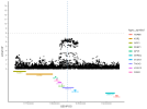
Chr12 contained three Tier 1 genes.

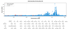
Image of the 2025 DecodeME results by @ME/CFS Science Blog:
1758826778973.png


********************
SUDS3 (Tier 1)

• Protein: SIN3A corepressor complex component, also known as SDS3. UniProt. GeneCards.
The allele that increases the risk of ME/CFS is associated with increased SUDS3 expression.7

• Molecular function: Regulatory protein that represses transcription and augments histone deacetylase activity of HDAC1 (UniProt). SUDS3 alters expression of the upstream kinase, ASK1, in the p38 MAPK pathway cascade (44).

• Cellular function: Chromatin remodeling and transcriptional regulation. Depletion of mouse Suds3 reveals an essential role in early lineage specification (45).

• Link to disease: Proposed to contribute to the microglial inflammatory response (44).

• Potential relevance to ME/CFS: Unclear, but potentially to neuroinflammation.

*******************

References
7. Johnson BS, Farkas D, El-Mergawy R, Adair JA, Elhance A, Eltobgy M, et al. Targeted degradation of extracellular mitochondrial aspartyl-tRNA synthetase modulates immune responses. Nat Commun. 2024 Dec 1;15(1).

44. Shen J, Lai W, Li Z, Zhu W, Bai X, Yang Z, et al. SDS3 regulates microglial inflammation by modulating the expression of the upstream kinase ASK1 in the p38 MAPK signaling pathway. Inflamm Res. 2024 Sep;73(9):1547–64.
(note SDS3 is an earlier name for SUDS3)

45. Zhang K, Dai X, Wallingford MC, Mager J. Depletion of Suds3 reveals an essential role in early lineage specification. Dev Biol. 2013 Jan 15;373(2):359–72.
 
Last edited:
No mentions of SUDS3 or SDS3 on the forum as yet, however

Other mentions of HDAC1 on the forum


HDAC1 was a significant hit in the Zhang study, no?

[Edit: ah sorry that was already mentioned above. I think it's interesting to see a few hits between studies on general epigenetic regulation, which might provide hints for findings in cultured cells, which are more likely to be reflective of epigenetic changes that persist in culture]
 
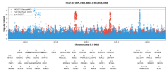
Copying @ME/CFS Science Blog's post on this region and TAOK3 here and also into the first post:
A gene that hasn't been disucssed much is TAOK3 on chromosome 12 (it wasn't a Tier 1 gene). It has been previously been associated with Lupus at around the same region as in DecodeME. The vertical dotted line in the graph below shows the location for the Lupus hit (12:118244946) with the SNP summary data from DecodeME.


1758826778973.png


The Lupus GWAS said this about it:
We also identified a missense variant in TAOK3 (the gene for tau kinase 3) as the top association signal in this locus. The risk allele (rs428073-T) substitutes the 47th amino acid of TAOK3 fromserine to asparagine (S47N), whose functional role remains unknown. S47 is located at the loop region between strands β2and β3, and the substitution should not change the overall strucure of the protein, despite being well conserved among orthologous proteins during evolutionary courses (SupplementaryFigure 10, on the Arthritis & Rheumatology website at https://onlinelibrary.wiley.com/doi/10.1002/art.42021). Taok3 plays animportant role in DNA damage–induced activation of the p38/MAPK14 stress-activated MAPK cascade. It enhances T cell receptor signaling by regulating its negative feedback by SH2domain–containing phosphatase 1 (44), and Taok3 deficiency in mice was found to cause defects in the development of marginalzone B cells but not follicular B cells (45).
Identification of Shared and Asian-Specific Loci for Systemic Lupus Erythematosus and Evidence for Roles of Type III Interferon Signaling and Lysosomal Function in the Disease: A Multi-Ancestral Genome-Wide Association Study - PubMed
In this study both shared and Asian-specific loci for SLE were identified, and functional annotation provided evidence of the involvement of increased type III IFN signaling and reduced lysosomal function in SLE.
pubmed.ncbi.nlm.nih.gov
 
"It enhances T cell receptor signaling by regulating its negative feedback by SH2domain–containing phosphatase 1 (44), and Taok3 deficiency in mice was found to cause defects in the development of marginalzone B cells but not follicular B cells (45)."

Intriguing. An influence on SHIP1 would make some sense. Marginal zone B cell behaviour is odd in lupus too.
 
Last edited:
Interesting, so it seems like this is another case where the final annotation for DecodeME was chosen based on high number of color tissues. The top SNP is within an intronic region in DecodeME, and seems to be quite a long deletion. The mutation in the lupus paper you posted was in a protein-coding region which might explain stronger effect of the mutation.

Also, interesting that 3 proteins in that whole region that looks to be in LD have well-established links to MAPK signaling (SUDS3, TAOK3, and PEBP1)
 
Posts moved from main DecodeME thread.
---------------------------------------------------------


it'd probably be very nice for both SLE and ME/CFS research to find a common pathway/gene.
Could you also check the region above TAOK3 on chromosome 12? It was found to be associated with lupus according to the GWAS catalog.
 
Last edited by a moderator:
Could you also check the region above TAOK3 on chromosome 12? It was found to be associated with lupus according to the GWAS catalog.
It doesn't look like it was significant in this study. And the Bentham 2015 paper doesn't mention any locus in this region. It mentions two other loci on chromosome 12, which can be seen if I zoom out really far. The ME/CFS TAOK3 locus is the left red tower. (Not all gene names shown.)
1761138225476.png

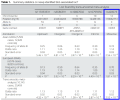
Looking at the lupus paper you're referring to, Wang et al, here's the data for that TAOK3 SNP (right side of table cut off):
image.psd(6).png

They got 1.81e-3 for the p value of that SNP when they only look at ~5000 European cases, which is close to the sample size in the Bentham study. This p value isn't very far off Bentham et al's result of p=1.6e-2 (from the summary statistics).

It's only when Wang et al combined the European cases with another ~5000 Asian cases that it passed the genome-wide significance threshold.
 
Could you also check the region above TAOK3 on chromosome 12? It was found to be associated with lupus according to the GWAS catalog.
That paper refers to a study/dataset from the same author from a year earlier: GCST011096. It has slightly fewer Asian cases, but it looks like the same European sample. So it is based on 8798 cases and 16470 controls from mixed ancestry.

I was able to download this data and plot it. And it does look like basically the same area for the locus in SLE and ME/CFS:
ME_CFS__DecodeME__SLE__Wang_2021__chr12:117494946-118994946.png

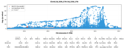
Also, this dataset demonstrates the benefit of multi-ancestry GWAS. Here's the chr6p locus when looking at the European cases from Bentham et al:


Here is the same area, but looking at the data when European and Asian cases were combined in Wang 2021:
1761147366692.png

Linkage disequilibrium patterns are different between ancestries, so if both populations share a common causal SNP, but this SNP is in LD with different non-important SNPs in the two populations, far fewer non-important SNPs will be significant, helping to narrow down where the actually causal variant is.

(Though the difference in shape isn't necessarily completely due to different LD. The two populations might not have identical causal variants.)

Same thing said in a paper:
Second, single-ancestry GWAS are hampered by the specific linkage disequilibrium (LD) structure in that ancestry, which could obscure the ability to effectively fine-map an associated locus. Multi-ancestry GWAS can improve fine-mapping resolution by leveraging the distinct LD structures in each ancestry
 
Last edited:
That paper refers to a study/dataset from the same author from a year earlier: GCST011096. It has slightly fewer Asian cases, but it looks like the same European sample. So it is based on 8798 cases and 16470 controls from mixed ancestry.

I was able to download this data and plot it. And it does look like basically the same area for the locus in SLE and ME/CFS:
ME_CFS__DecodeME__SLE__Wang_2021__chr12:117494946-118994946.png
I'll note that from my amateur attempt to try to determine if both studies share a causal variant at this locus, it doesn't seem like it's the case that they do.

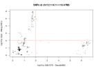
First of all, when looking only at the region that includes this locus, chr12:118110000-118470000, there are 344 SNPs that are included in both datasets. Of these, 58 SNPs have p<.01 in ME/CFS and 78 SNPs have p<.01 in SLE. But only a single SNP has p<.01 in both (and just barely in ME/CFS).

Here's the same analysis as a plot. If there were SNPs that were very significant in both GWAS, they would be in the upper right area, but this area is basically empty.
snp_ps_chr12.png

I also tried to use the coloc function to test whether they share a common variant. It seems fairly straightforward to run, but it's my first time doing it, so I might have done something wrong. I'll include resources I read about coloc and the code I used at the end of the post.

This was the result:
P = probabilty

H0: 0.00036 - P(no signal in either study)
H1: 0.051 - P(signal only in ME/CFS)
H2: 0.0066 - P(signal only in SLE)
H3: 0.93 - P(signal in both studies, but they do not share a causal variant)
H4: 0.0072 - P(signal in both studies, and they share a causal variant)
According to this, there is only a less than 1% chance that the two studies have a shared causal variant in this region.

I also think, but am not sure, that the studies should have the same ancestries for coloc, so that might make this result not reliable since the ancestries don't match. In which case, I'd say the above part about lack of shared significant SNPs is still probably good evidence against them sharing a causal variant.

Code:
library(coloc)

gwas1 <- read.table('~/Projects/mecfs/mecfs_science/studies/Ponting 2025 - DecodeME/Data/filtered_regenie_file.txt.gz', header=TRUE)
gwas1$P <- 10**-gwas1$LOG10P

gwas2 <- read.table('~/Projects/mecfs/mecfs_science/studies/Ponting 2025 - DecodeME/Data/Other Studies/SLE/Wang 2021/wang2021_harmon_id-added_38.tsv.gz', header=TRUE)
gwas2$log10p_value <- -log10(gwas2$p_value)

chr <- 12
region_start <- 118110000
region_end <- 118470000

gwas1_region <- gwas1[gwas1$CHROM == chr &
                        gwas1$GENPOS >= region_start &
                        gwas1$GENPOS <= region_end, ]

gwas2_region <- gwas2[gwas2$chromosome == chr &
                        gwas2$base_pair_location >= region_start &
                        gwas2$base_pair_location <= region_end, ]

intersection <- merge(gwas1_region, gwas2_region,
                      by.x = "ID", by.y = "id")

gwas1_sig <- sum(intersection$P < .01)
gwas2_sig <- sum(intersection$p_value < .01)

sig_both <- sum(intersection$P < .01 & intersection$p_value < .01)
cat("Number of overlapping SNPs:", nrow(intersection), "\n")
cat("Overlapping SNPs with p<.01 in GWAS 1:", gwas1_sig, "\n")
cat("Overlapping SNPs with p<.01 in GWAS 2:", gwas2_sig, "\n")
cat("Overlapping SNPs with p<.01 in both GWAS:", sig_both, "\n")

plot(intersection$LOG10P, intersection$log10p_value,
     xlab = "-log10 p (ME/CFS - DecodeME)",
     ylab = "-log10 p (SLE - Wang 2021)",
     main = sprintf("SNPs at chr12:%.2f-%.2fMb", region_start/1e6, region_end/1e6),
     pch = 19,
     cex = 0.5,
     col = rgb(0, 0, 0, 0.3)
)

abline(h = -log10(0.01), col = "red", lty = 2)
abline(v = -log10(0.01), col = "red", lty = 2)

# Coloc

s_gwas1 <- gwas1_region$N_CASES[1] / gwas1_region$N[1]
s_gwas2 <- 8798/(8798+16470)

gwas1_list <- list(
  beta = gwas1_region$BETA,
  varbeta = gwas1_region$SE^2,
  snp = gwas1_region$ID,
  position = gwas1_region$GENPOS,
  type = "cc",
  s = s_gwas1,
  N = gwas1_region$N[1]
)

gwas2_list <- list(
  beta = gwas2_region$beta,
  varbeta = gwas2_region$standard_error^2,
  snp = gwas2_region$id,
  position = gwas2_region$base_pair_location,
  type = "cc",
  s = s_gwas2,
  N = 16470
)

check_dataset(gwas1_list)
check_dataset(gwas2_list)

coloc_result <- coloc.abf(dataset1 = gwas1_list,
                          dataset2 = gwas2_list)

format(coloc_result$summary, scientific = FALSE)

sensitivity(coloc_result, "H4 > 0.5")
- https://chr1swallace.github.io/coloc/articles/a02_data.html
- https://github.com/orlandocox/How-to-conduct-Post-GWAS-analysis/blob/main/5.0 Colocalization Analysis.md
 
First of all, when looking only at the region that includes this locus, chr12:118110000-118470000, there are 344 SNPs that are included in both datasets. Of these, 58 SNPs have p<.01 in ME/CFS and 78 SNPs have p<.01 in SLE. But only a single SNP has p<.01 in both (and just barely in ME/CFS).

This seems a very useful way of showing that two risk packets are definitely not the same. I forget which candidate gene this relates to for ME/CFS?

One might guess that for any given gene there might be a risk packet, or group of them that will track together for any given disease, for more and another one for less? If it is more complicated I am not sure what that should be interpreted as.
 
I forget which candidate gene this relates to for ME/CFS?
This is the TAOK3/SUDS3 region.

One might guess that for any given gene there might be a risk packet, or group of them that will track together for any given disease, for more and another one for less? If it is more complicated I am not sure what that should be interpreted as.
The GTEx project shows that variants alter expression differently in different cell types/organs. So I think that's possible.

For example, maybe the ME/CFS variant increases TAOK3 in the liver while the SLE variant decreases TAOK3 in the skin, or even alters the shape of the TAOK3 protein directly (the lupus study @ME/CFS Science Blog posted suggested this latter option).
 
Back
Top Bottom