Plasma proteomic profiling suggests an association between antigen driven clonal B cell expansion and ME/CFS, 2020, Lipkin et al

Andy

Retired committee member
Myalgic Encephalomyelitis/Chronic Fatigue Syndrome (ME/CFS) is an unexplained chronic, debilitating illness characterized by fatigue, sleep disturbances, cognitive dysfunction, orthostatic intolerance and gastrointestinal problems.

Using ultra performance liquid chromatography-tandem mass spectrometry (UPLC-MS/MS), we analyzed the plasma proteomes of 39 ME/CFS patients and 41 healthy controls. Logistic regression models, with both linear and quadratic terms of the protein levels as independent variables, revealed a significant association between ME/CFS and the immunoglobulin heavy variable (IGHV) region 3-23/30.

Stratifying the ME/CFS group based on self-reported irritable bowel syndrome (sr-IBS) status revealed a significant quadratic effect of immunoglobulin lambda constant region 7 on its association with ME/CFS with sr-IBS whilst IGHV3-23/30 and immunoglobulin kappa variable region 3–11 were significantly associated with ME/CFS without sr-IBS.

In addition, we were able to predict ME/CFS status with a high degree of accuracy (AUC = 0.774–0.838) using a panel of proteins selected by 3 different machine learning algorithms: Lasso, Random Forests, and XGBoost. These algorithms also identified proteomic profiles that predicted the status of ME/CFS patients with sr-IBS (AUC = 0.806–0.846) and ME/CFS without sr-IBS (AUC = 0.754–0.780).

Our findings are consistent with a significant association of ME/CFS with immune dysregulation and highlight the potential use of the plasma proteome as a source of biomarkers for disease.
Open access, https://journals.plos.org/plosone/article?id=10.1371/journal.pone.0236148
 
We pursued proteomic analyses of plasma from subjects with ME/CFS with the objectives of obtaining insight into the pathogenesis of ME/CFS and finding biomarkers for the disease. Using models with only linear terms, we did not identify significant differences between cases and controls following FWER correction. However, using a model that included both linear and quadratic terms we found a significant association between ME/CFS and IGHV3-23/30. The association with quadratic effect revealed both a positive and negative correlation relationship between levels of IGHV3-23/30 and ME/CFS. Whilst 12 patients had extremely high levels (>100,000) of IGHV3-23/30, only 3 patients had extremely low levels (<25,000) of this protein. IGHV3-23 is one of the most commonly used heavy variable regions in the human immunoglobulin (Ig) repertoire [30]. Its usage has been linked with non-Hodgkin lymphomas (NHL) such as chronic lymphoid leukemia (CLL) [3134], mantle cell lymphoma (MCL) [35], splenic marginal zone lymphoma (MZL) [36, 37], Waldenström’s macroglobulinemia [38], and follicular lymphoma (FL) [39, 40]. Disease progression in these B-cell malignancies is driven by chronic stimulation from either microbial or auto-antigens. Interruption of B-cell signaling through use of kinase inhibitors has been shown to have therapeutic benefit in some patients with MZL, CLL, FL, and MCL [41, 42]. Increased IGHV3-23 usage is also reported in anti-myelin associated glycoprotein neuropathy and in monoclonal gammopathy of undetermined significance [43], a disorder that may progress to malignant lymphoproliferative disease [44]. We speculate that, at least in a subset of ME/CFS subjects, an increased level of IGHV3-23 may be due to antigen driven clonal expansion, and that these patients might benefit from identification of the antigen driving B-cell receptor signaling or kinase inhibitors that interrupt signaling. In this context, we also note that ME/CFS has been linked with an increased risk of developing MZL and other NHL [45].

Sounds like they interpret this as an autoimmune problem.

But how plausible is B cell autoimmunity after the negative Rituximab trial?
 
Sounds like they interpret this as an autoimmune problem.

But how plausible is B cell autoimmunity after the negative Rituximab trial?


I've not read the whole paper yet, but that paragraph from the discussion section says:
Disease progression in these B-cell malignancies is driven by chronic stimulation from either microbial or auto-antigens. Interruption of B-cell signaling through use of kinase inhibitors has been shown to have therapeutic benefit in some patients with MZL, CLL, FL, and MCL [41, 42].

I would take that to mean "either microbial antigens or auto-antigens," so not necessarily auto-immune.

Microbes could be many things:
Microorganisms can be bacteria, fungi, archaea or protists. The term microorganisms does not include viruses and prions, which are generally classified as non-living.
https://www.uib.no/en/geobio/56846/what-are-microorganisms

It could be a chronically "present" microorganism that the immune system can't get rid of. One source for that that might be microbes in the gut somehow crossing a boundary which brings them to the attention of the immune system. It would be a small enough "infection" for the immune system to handle, but it would be "chronic" so long as the cause of the "leak" was not addressed.
 
Last edited:
Im going to try to explain what this study found.

First, we need to be familiar with the structure of antibody. An antibody is an immunoglobulin (Ig) that can come in several 
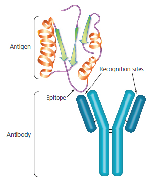
classes (G, A, M, D and E). This is the prototypical antibody:

antibody.jpg


Notice the red part. This is called the heavy chain. The blue part is called the light chain. Each light chain is connected to a heavy chain via disulphide bonds and the heavy chains themselves are connected via disulphide bonds, giving a macromolecule with two heavy chains and two light chains.

An antibody recognises and covalently binds to a specific "thing". This "thing" is called an antigen and can be many things: proteins, LPS, even hormones. In order for the body to recognise a wide variety of antigens, it makes sense to have many different antibodies. This is achieved by having a constant region, that determines the common characteristics of all antibodies (for example, the class), and a variable region. This variable region is the one that binds to antigen, and comes in many different shapes.

antibody-antigen-interaction.jpg


This is how an antibody binds. In this case, the antigen is a protein, and the small part that the antibody recognises is called epitope. An epitope is a part of the antigen, and the same antigen can have different epitopes. For example a big protein has different epitopes, each one corresponding to some different amino acid sequence.

It is in the best interest of the body to recognise a wide variety of different antigens, in order to have a broad spectrum of defence, but it is impractical to have thousands or tens of thousands of genes, each coding for a different variable region.

So how does the body generate the observed antibody diversity? By a complex process called somatic recombination. In this process, different genes are joined together to form variable regions. Because of the many different ways the genes can be joined, the diversity increases exponentially while using comparatively few genes.

diversity-3-w-padding-01.png


This is how somatic recombination works. For the Variable part of the heavy chain, a particular Variable gene is chosen. This gene is combined with a Diversity gene, and the two combine with a Join gene. This is called V(D)J recombination, and the VDJ sequence is finally joined to a Constant gene, that encodes the constant region.

For the light chain, only Variable and Join genes combine to form the Variable part, therefore the light chain has less diversity.

Once the heavy chain and light chain are transcribed and translated, the protein products join to form a functioning antibody.

The antibody producing cells are called B cells, and each B cell produces only one kind of antibody (though exceptions occur). Over the lifetime of a B cell however, it can class switch. Changing the Constant region of the heavy chain to a different class.

A B cell is first inactive, generating antibody and using it as a membrane bound receptor, the B Cell Receptor (BCR). When the membrane receptor recognises and binds to antigen, the B cell becomes activated. It processes the encountered antigen and presents it to T helper cells. If the T cell also recognises it, it "authorises" the B cell to activate fully.

The B cell rapidly replicates, migrates to the blood and tissues, and starts secreting the antibody. This process of rapid replication upon activation is called clonal expansion, and most of the clonal expanded B cells produce the same antibody, although there are special mechanisms to subtly change the variable region of a few B cells to generate more potent antibodies.

OSC_Microbio_18_04_BCellact.jpg


The image above is an example of clonal expansion.

Now, what did Lipkin find? He found a particular protein elevated in the blood. This protein is called IGHV3-23, this stands for Immune Globulin Heavy chain Variable 3 -23. This gene is one of the Variable genes that we talked bout earlier.

The reasoning is that because this protein is elevated in blood, some B cell has become activated and is producing large quantities of one particular antibody that contains IGHV3-23, therefore generating large amounts of this IGHV3-23.

B cell activation is suggestive of chronic infection or autoimmunity. Which one is it? I have no idea:)
 
Last edited:
I am not sure but I think this is on topic. Imaging of my right neck lymph node in 2009 showed a diameter of 1,74 cm. According to this article
In general, lymph nodes are considered abnormal if their diameter exceeds one cm.

It has always been this way and sometimes it's sore. A few other lymph nodes are also not normal.

PS: forgot the link https://www.ncbi.nlm.nih.gov/pmc/articles/PMC3993046/
 
Last edited:
The main message I get from this is that there were no differences between ME/CFSand controls on a straightforward comparison of levels of expression of VH genes (I have always been familiar with the term VH, rather than IGHV). The difference they claim appears with a mathematical transformation that I do not understand the significance of.

I am also a bit worried about the method of selecting cases from several tertiary referral centres.At lets some of these centres have a reputation for being interested in e.g. EBV and may get cases referred because of unusual immune profiles in the first place.

Different proportions in usage of VH (IGHV) genes may have all sorts of causes. Skewed usage is not particularly easy to detect in autoimmune disease so probably has nothing much to do with any particular antigen. Certain genes like VH4-34 are over used in several conditions but this may not be a reflection of the antigens being handled.

The link to lymphoma if anything suggest to me that if this result is a real effect it may indicate that a proportion of PWME have some sort of pre-neoplastic B cell expansion perhaps analogous to MGUS (monoclonal gammopathy of unknown significance). Although there is some evidence for antigen drive in some neoplastic and pre-neoplastic states the majority look to be independent of antigen drive as far as I am aware.

So I don't think this points particularly to autoimmunity or to persistent drive from a microbial antigen although both are possible. The negative rituximab trial does not exclude autoimmune involvement because only a proportion of autoimmune diseases respond. Response is dependent on the condition being mediated by short lived plasma cells, which is true for RA but probably not for e.g. scleroderma.
 
I've not read the whole paper yet, but that paragraph from the discussion section says:


I would take that to mean "either microbial antigens or auto-antigens," so not necessarily auto-immune.

Microbes could be many things:


It could be a chronically "present" microorganism that the immune system can't get rid of. One source for that that might be microbes in the gut somehow crossing a boundary which brings them to the attention of the immune system. It would be a small enough "infection" for the immune system to handle, but it would be "chronic" so long as the cause of the "leak" was not addressed.
So the recently found genetic glitch that significantly affects mucosal areas ( nose / lungs/ gut) may offer a route ?
 
The main message I get from this is that there were no differences between ME/CFSand controls on a straightforward comparison of levels of expression of VH genes (I have always been familiar with the term VH, rather than IGHV). The difference they claim appears with a mathematical transformation that I do not understand the significance of.

Yes, this was my impression as well after looking at the table of raw results. The difference in IGVH3-23/30 is rather small, and in the human body most significant interactions are first-order, though not always linear, so the use of a second-order transform does not easily have "physical" meaning.

These kinds of results are so common in ME/CFS research that looks at the immune system and at bioenergetics, that I would take them as a sign that its time to look at other systems, particularly the CNS.
 
A Proteomics Study from the Center: Searching for the Criminal
..
What did the latest study from the Center show? Basically, two things:

  • There appears to be a distinctive “signature” of a small group of proteins that distinguishes people with ME/CFS from healthy people;
  • The proteins involved in that “signature” are primarily involved in the immune response—particularly the response of immune cells called B cells—to infections, and the response seen in autoimmune diseases.
https://cfsformecfs.org/2020/07/28/a-proteomics-study-from-the-center-searching-for-the-criminal/
 
I was please to read this section. It reinforces that @Chris Ponting who has a Phd researher looking at Tcell clonal expansion and is collaborating with a researcher at Oxford to look at B cell clonal expansion is on the right track to look for clues......
Also, if further studies can identify the antigens that the activated immune system is activated by, these results may reveal underlying toxins, infectious agents, or other foreign molecules that can themselves be targeted .
 
I was please to read this section. It reinforces that @Chris Ponting who has a Phd researher looking at Tcell clonal expansion and is collaborating with a researcher at Oxford to look at B cell clonal expansion is on the right track to look for clues......

There was also a collegue of Chris Pointing (from Edinburgh) at the CMRC conference who was interested in B cells and starting to think about ME.
 
What I liked about this was the stratification of the ME/CFS patients into subgroups based on symptoms with corresponding significant differences in their proteome.

This was recognised as a method without discussing the implications of significant differences detected using this method in the abstract but is imho potential progress towards effective subtyping, which I believe will be helpful in selecting homogenous patient groups and distinguishing and characterising different conditions with similar presentations as well as different stages of progression in identified subtypes.

At this point I dont think we can say whether PWME w/o IBS are a different subtype or a difference progression / cycle stage cf PWME w IBS. It would be a significant advance if we could find ways to distinguish subtypes by reliable empirical methods and then use these to define experimental cohorts imho.

I had to watch a video to understand what a quadratic analysis is but it looks good to me if the square of a measure (quadratic plot) allows recognition of statistical significance when a plot of the measure itself (linear plot) does not. It indicates something about the delicate biological processes regulating expression of proteins which are often subject to homeostatic regulation which could credibly make linear analysis less sensitive to state changes than quadratic imho.
 
A point raised by Jo Cambridge that I did not mention is that a shift in VH (or IgHV) gene usage is not an indicator of clonal expansion as such. To identify clones you need to sequence the antigen binding section of the antibody (CDR or complementarity determining region).
 
I have an advisory role on the Columbia U ME/CFS center. They wanted to know if folks had any questions for their researchers on their study "Plasma proteomic profiling suggests an association between antigen driven clonal B cell expansion and ME/CFS, 2020, Lipkin et al"

If you have any questions, I will forward them and they will try to have their researchers answer them on their web site.


Questions I see so far.

1) Can you please explain the biological significance of the quadratic transformation of the data?

2) To demonstrate B cell clonal expansion, do you need to sequence the antigen binding section of the antibody (CDR or complementarity determining region)?
 
Last edited by a moderator:
Back
Top