Microvascular Dysfunction and Basal Membrane Thickening in Skeletal Muscle in ME/CFS and Post-COVID, 2025, Slaghekke et al

Siebe

Established Member
I'm copy-pasting a Twitter thread that I wrote about a poster presentation by Slaghekke et al. (Wüst team). I've added an extra sentence here and there (e.g. on Poiseille's law) to explain a bit more.

You can read a general take by ME/CFS Science in the tweet below.

Poster: https://mecfs-research.org/wp-content/uploads/2025/04/Anouk-Slaghekke_Poster_Conference_2025.pdf
Presentation:
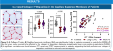
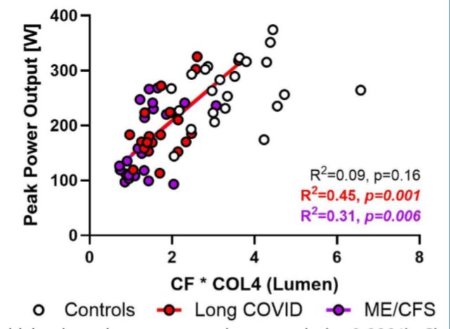
1000049726.pngWhat's interesting about these findings, is that they had moderately strong correlation with CPET performance! That's in addition to there being a clean separation between healthy controls vs. LC* & ME

I find these findings really exciting!

Some thoughts & caveats


Unroll available on Thread Reader

Image

First, a methodology question

The x-axis appears to be a composite measure of 'CF * COL4 (Lumen)'

(Lumen is the internal space of the capillary, through which the blood flows)

In the caption it's called 'CF*Lumen'

I don't know what CF means. Capillary flow? Collagen fiber? Image

Also, you can create all kinds of composite measures, so this increases the risk of p-hacking.

But if we leave that aside and assume that there's a benign reason for it, we're left with some fascinating results.. because these findings aren't unique to LC/ME

Scleromyositis is a condition where there's significant thickening of the basement membrane, as well as high amounts (~two-thirds) of BM 'reduplication' (layers) Image
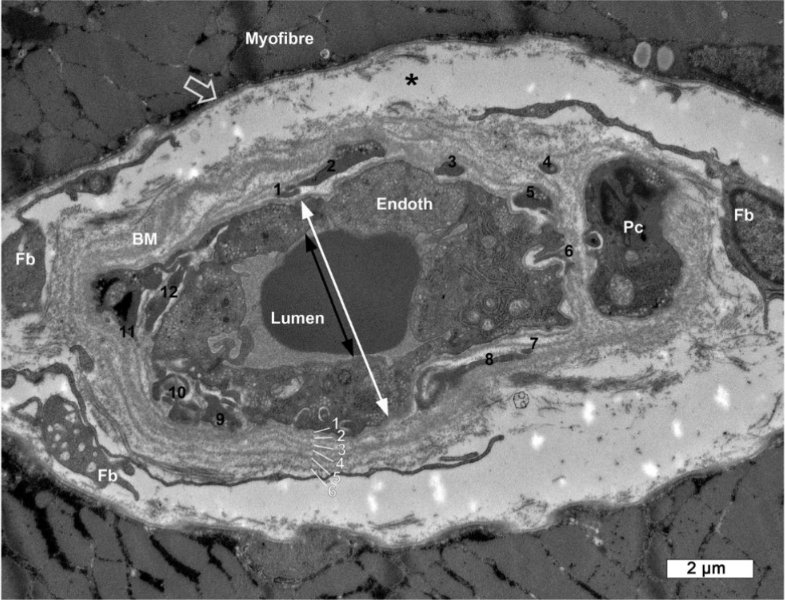
Image of a capillary in Scleromyositis. You can see the 6 layers of the basement membrane (should normally be just one)

Now, Slaghekke doesn't note the frequency of reduplication in LC/ME samples, and I can't find comparable BM measurements in scleromyositis.

But we can assume it's a worse amount of BM thickening, as well as that it's systemic (including the lungs). Image
Image of reduplication in ME/CFS, from the poster

We don't yet know if it's systemic in LC/ME or only in muscle capillaries.

Regarding the lumen diameter, I can't easily find data for Scleromyositis, so it's hard to compare. (This matters, as I discuss later)

Now the interesting question: how do people with scleromyositis react to exercise? Do they get PEM?

They do have a 'disproportional response to exercise', getting easily fatigued. This is not just due to capillary BM thickening in muscles, but also in the lungs and at the heart.

However, scleromyositis patients do improve with exercise over time! No PEM. This suggests ME/CFS has more going on.

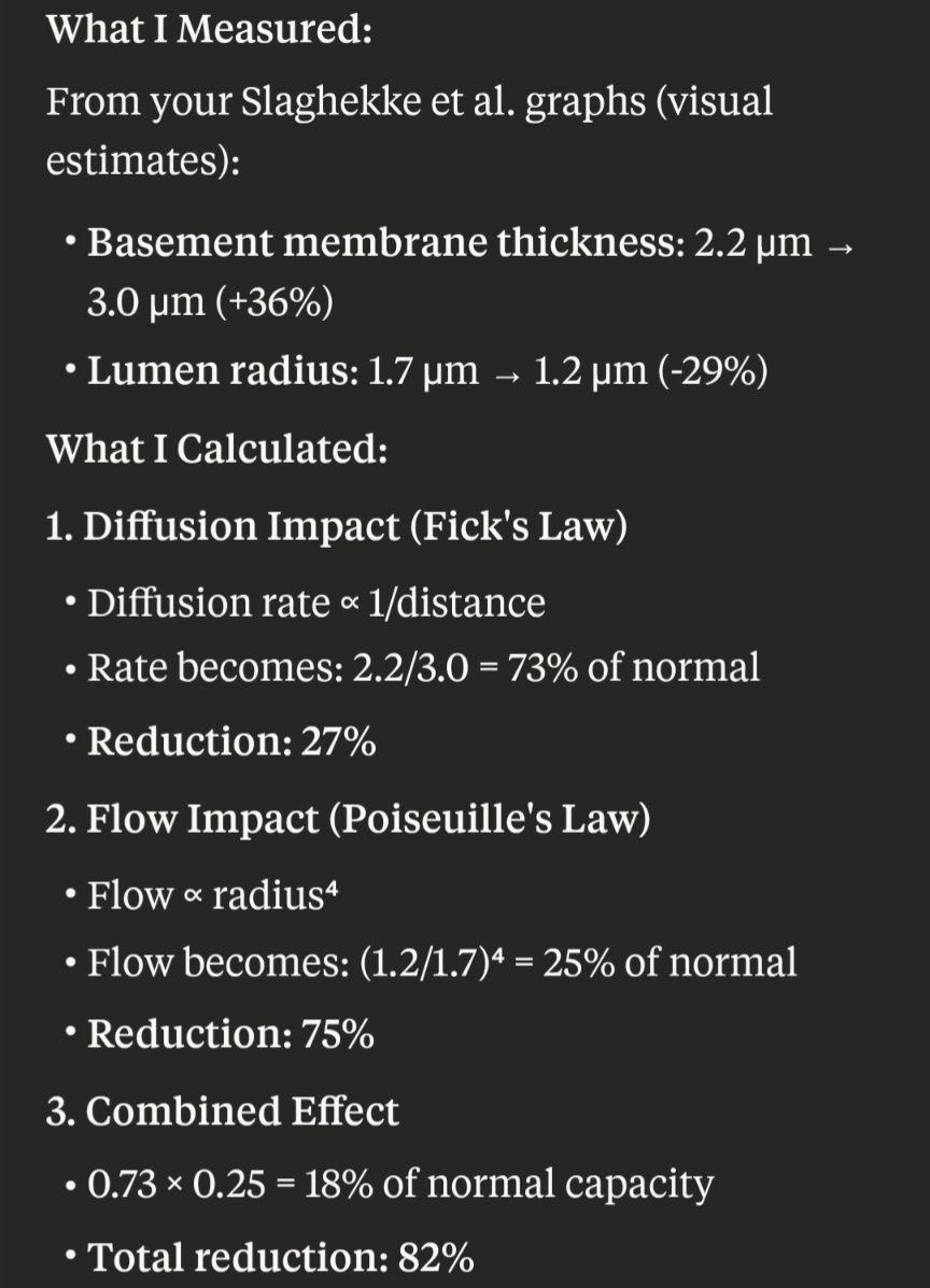
Still, it has an effect on 1-day CPET performance. I asked Claude (AI) to calculate how much these variables would affect oxygen supply, and was pretty shocked!

The diffusion would be reduced by 25-35% (thicker layer to cross) Image
1000049782.webp
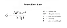
Flow however is much more severely impacted by reduced diameter (see the r⁴ term). A 29% reduced diameter leads to a 75% reduced blood flow through the capillaries! (Reduced volume at reduced speeds) I'm not familiar with this, maybe this is compensated somewhat by higher pressure?

Together, these would lead to 81-84% reduction in capacity! Image
(I don't know how this compares to scleromyositis, because of the unknown lumen space. I'm on completely unfamiliar terrain, but I think lumen space might be significantly wider and more variable in Scleromyositis. This would imply less flow issues and more oxygen delivery)

Now, 81-84% doesn't quite match the observed reductions in power output, which looks more like 30-60%.

This could be due to compensatory mechanisms, such as anaerobic respiration. Image
Nonetheless, it would be great if we can resolve this, especially by increasing the lumen space, which would have the largest effect on capacity

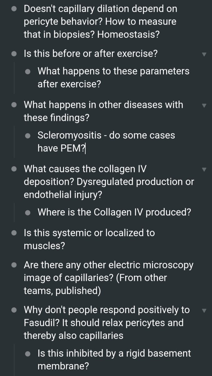
1 drug that would normally help to widen capillaries is Fasudil, which works by relaxing the pericytes attached to endothelial cells..

Well, I happen to know of 10+ people who took Fasudil. And iirc, no response.

How could that be? I don't know. Perhaps the basement membrane is stiff enough to keep the capillaries' lumen compressed regardless of pericyte relaxation?

This all leaves me with a bunch of questions, like all good research does. I hope we see a replication attempt soon, as well as highly needed follow-up research. I'm excited! Image
- What would happen if we let people do a CPET in a high-oxygen setting? HBOT could give up to 2x the oxygen (2.4ATA, my calculations), which wouldn't be enough to compensate for 5x capacity loss, but might still be interesting?
 
Last edited by a moderator:
As you say there's a lot of possibility for p-hacking when you combine different measures together (and there additionally appears to be no correction here) and as you say, the story doesn't sensibly add up because ME/CFS isn't just a story about "acute failure to exert and getting better with more training". I'm also not quite sure why the correlation to a one day CPET result would be interesting, because as far as I know one day CPETs tell us nothing about ME/CFS and Long-Covid, especially when the correlation reported here is to peak power output, not something like reduction in VO2 at peak in the second test.

This group has previously shown that they are very aware of patient selection, including looking at activity levels in controls, but without any data one should probably be cautious in overinterpreting findings which can be driven by different activity patterns (lower capillarization, lower peak power output ect).

I wonder about the NIRS data. If the story if supposed to be about lower O_2 in muscles why have other studies which have used NIRS (I think by now there's a handful of studies using NIRS in LC and/or ME/CFS) not shown anything or what explains inconsistent results?

What would happen if we let people do a CPET in a high-oxygen setting? HBOT could give up to 2x the oxygen (2.4ATA, my calculations), which wouldn't be enough to compensate for 5x capacity loss, but might still be interesting?
Wouldn't it be sufficient to just give patients an EPO drug like those that have been trialled in ME/CFS?
 
Wouldn't it be sufficient to just give patients an EPO drug like those that have been trialled in ME/CFS?
No. That would only increase oxygen content by 25%, but also increase viscosity due to higher hematocrit ratio which would reduce flow by more than 25%. In healthy people, that can be offset by capillary dilation, but this may not be possible for ME/CFS
 
I wonder about the NIRS data. If the story if supposed to be about lower O_2 in muscles why have other studies which have used NIRS (I think by now there's a handful of studies using NIRS in LC and/or ME/CFS) not shown anything or what explains inconsistent results?
I looked into the existing studies, and I don't think any results are particularly useful:

ME/CFS Peripheral NIRS:
- McCully series (1999, 2003, 2004) - dismissed due to Fukuda criteria
- Miller et al. (2015) - differences disappeared when work output matched between groups
- Walitt et al. (2024) - no differences in quadriceps oxygen saturation during maximal CPET. However, n=8 ME/CFS patients and 9 healthy volunteers.

Long COVID Peripheral NIRS:
- Seifart et al. (2023) - confounded by comorbidities (high rates of obesity, hypertension)
- Goulart et al. (2024) - no controls, obesity confound

Not NIRS but did find impaired oxygen extraction in subset:
Systrom invasive CPET (2021) - good methodology, clear findings
Thanks for looking into it.

I suppose one can dismiss studies using Fukuda criteria in the context of ME/CFS research, but I think it's not quite clear whether one can dismiss them in the context seen here (the authors seem to suggest that these ME/CFS and Long-Covid patients look similar, but everybody meeting Fukuda criteria will meet Long-Covid criteria, but it isn't quite clear whether those people would meet the criteria to be a Long-Covid patient in this study). It seems that the authors here also report that there are no differences in NIRS during exercise and that NIRS differences occur only post-exercise, which might explain previously negative findings.

In either case I think anybody will have a hard time to explain ME/CFS with differences in muscle oxygenation post exercise (the differences reported between controls and patients here is likely far less than between controls and athletes), when many patients report brain fog, hypersenstivity to sound and the inability to walk around the block. One may argue that it might only be "part of the picture" but then the more likely scenario is always that it is not be part of the picture.

Will be interesting to see how they managed to account for differing activity profiles and how that was accounted for in the different analyses (especially when some of the measurements should not be as activity dependent as others). Let's hope they will eventually get to present all the data not just a few pictures and p-values.
 
I would be cautious about thdse findings. The only thing I can decipher from figure 3A is that the MECFS sample has major ice artifact and the control doesn't. I am not sure that measuring basement membrane and lumens give us reliable information about the physiological state (as has been raised above).

I also don't understand why these changes would produce symptoms of MECFS. As noted before, starvation of muscles of nutrients and oxygen produces claudication pain, not what you get in MECFS.
 
As noted before, starvation of muscles of nutrients and oxygen produces claudication pain, not what you get in MECFS.
I have no investment in muscle studies, but I have spoken to several moderate-severe people with ME/CFS (CCC, diagnosed by a CCC author) who used to be athletes and describe their pain as being the "lactic acid build up" that they were used to as active athletes, which I interpret as meaning claudication pain.
 
I have no investment in muscle studies, but I have spoken to several moderate-severe people with ME/CFS (CCC, diagnosed by a CCC author) who used to be athletes and describe their pain as being the "lactic acid build up" that they were used to as active athletes, which I interpret as meaning claudication pain.
There is quite a bit of confusion over this and it may need detailed thinking through. I think the athletes' lactic acid story is a popular idea that may not have a precise basis. I have heard it said that lactate is not actually the cause for feeling "the wall" at twenty metres before the tape.

My own experience is of muscles refusing to respond but not specifically pain. I suspect claudication is different. It sounds very painful and what I would expect from ischaemia (no oxygen) rather than lactate excess.

Could impairment of capillary permeability lead to lactate build up without oxygen deprivation? Some others might have better info on this.
 
I would be cautious about thdse findings. The only thing I can decipher from figure 3A is that the MECFS sample has major ice artifact and the control doesn't. I am not sure that measuring basement membrane and lumens give us reliable information about the physiological state (as has been raised above).

I also don't understand why these changes would produce symptoms of MECFS. As noted before, starvation of muscles of nutrients and oxygen produces claudication pain, not what you get in MECFS.
I would be very surprised if you find many severe or even moderate-severe ME/CFS patient without (some form of claudication) muscle pain - especially with overexertion.

It's certainly not (exclusively) knowing that I am getting worse afterwards or 'central fatigue' that keeps me from moving. I do think the CNS is the main culprit as even cognitive exertion can cause claudication pain in the thighs (a very well known and often described and unfortunately ignored phenomenon in severe patients), but I suspect there is something else going on that's related to cellular hypoxia in the periphery.
 
Claudication is not just muscle pain. It is doecifically recognized as coming on after a fixed amount of exertion and even more importantly fissppearing with a fixed period of rest. I have never heard of the latter in accounts from lpeople with MECFS .
 
Claudication is not just muscle pain. It is doecifically recognized as coming on after a fixed amount of exertion and even more importantly fissppearing with a fixed period of rest. I have never heard of the latter in accounts from lpeople with MECFS .

Sounds a lot like what many patients describe, imo. That said, I doubt what happens in ME/CFS is equivalent to 'classical claudication' - which I believe is mostly related to a purely arterial pathology?

That's of course compete speculation, but I think the 'claudication' pain in ME/CFS is potentially (and mostly) downstream of central hypoxia.
 
There is quite a bit of confusion over this and it may need detailed thinking through. I think the athletes' lactic acid story is a popular idea that may not have a precise basis. I have heard it said that lactate is not actually the cause for feeling "the wall" at twenty metres before the tape.

My own experience is of muscles refusing to respond but not specifically pain. I suspect claudication is different. It sounds very painful and what I would expect from ischaemia (no oxygen) rather than lactate excess.

Could impairment of capillary permeability lead to lactate build up without oxygen deprivation? Some others might have better info on this.
I liked this guy's explanation:

Didn't somebody find that our inorganic phosphate was a bit high recently? I remember thinking, huh, that's actually one of the few things that has shown up on my blood tests a few times.
 
Hm. The story has been that lactic acid is cleared so quickly it couldn't cause muscle pain/fatigue. But what about in ME/CFS, where it is not cleared quickly? See Jones et al. 2011 https://pubmed.ncbi.nlm.nih.gov/21749371/

"The CFS patients in the normal PCr depletion group also exhibited significant prolongation (almost 4-fold) of the time taken for pH to recover to baseline following exercise (Fig. 5a) suggesting the absence of any form of compensatory response to mitigate against the effects of excessive exercise-induced acidosis in muscle. The net effect is sustained and significant acidosis in muscle during and following exercise which is likely to have a significant impact on muscle function and contribute significantly to the expression of fatigue. A key factor in the suboptimal recovery response to muscle acidosis is the previously described delay [18] in achieving maximum proton excretion in response to acidosis, an observation confirmed in this study (Fig. 5b)."
 
Or is the problem not the lactic acid, but what the lactic acid signals - if we can't clear it, and it signals to the immune system to come do something, then do we keep signalling to the immune system that it needs to fix something and that makes us feel bad?
 
That's of course compete speculation, but I think the 'claudication' pain in ME/CFS is potentially (and mostly) downstream of central hypoxia.

If there was central hypoxia then people with ME/CFS would be blue, as people with central hypoxia are, and their sats would show it. Moreover, it would make the vessel thickness theory redundant.

I just don't see any a priori reason to think there is an oxygen problem. It seems to be something that lab scientists have latched on to without making sure it fits what they are actually trying to exlain clinically.
 
Also, I don't see why, if the problem is thick capillaries, maybe not letting nutrients in, there should be much lactate in people with ME/CFS trying to do simple tasks and finding it hard. Lactate is supposed to build up with maximal muscle usage.

What we need are some basic muscle biologists outside the ME/CFS and sports medicine fields who understand these things and their relation to clinical presentations. The ones I have met over the years all say that there is no evidence for a problem with things like oxygen usage and ATP production in muscle in ME/CFS.
 
As a mostly bedbound person with post COVID ME/CFS, I would say it feels like claudication in my thighs. My walking distance gradually became very short. I need to walk very slowly and sometimes I want to stop due to pain and weakness in my thighs (n=1)

David Systrom has seen quite a few iCPETs in patients with ME/CFS and LC. I believe poor oxygen extraction (low arteriovenous difference) should be taken seriously and should be explained. There is a range of possible reasons and one of them is capillary basement membrane thickening.
 
Last edited:
I have no investment in muscle studies, but I have spoken to several moderate-severe people with ME/CFS (CCC, diagnosed by a CCC author) who used to be athletes and describe their pain as being the "lactic acid build up" that they were used to as active athletes, which I interpret as meaning claudication pain.

I agree, this I believe is common in more moderate to severe patients. I am one, and from early in the disease onset I had a progressive worsening of this experience with just short walks. I wouldn't describe it as cramping, but more like the limb becoming more stiff or 'rigid', you progressively walked slower until it became too difficult and painful to continue. Rest was the ONLY thing that could repair it. But even during the first hours of rest, the legs remained very uncomfortable and burning. Jokingly, I referred to this experience when walking as rigor mortis setting in, but all jokes aside, that's precisely what it feels like.

I am however not convinced this is lactic acid build up. I think it is just poor blood flow and lack of oxygen, which is as I understand the definition of claudication, which improves with rest.

My organic acids test revealed low lactate and undetectable pyruvate. This is very common with ME/CFS patients: low pyruvate AND low lactate, and the research supports this finding. So the substrate delivery to pyruvate / lactate pathways are inhibited. I believe this is the active PKM2 enzyme, but that's another story. So, I'm not confident lactic acid buildup / lactate is a problem in ME/CFS, instead I think glycolytic metabolism is being rerouted through the PPP for NADPH production attempting to maintain redox homeostasis, this would be upregulated with exercise I imagine.

Furthermore, when I had a venous blood gases test (twice in fact), the results showed I had (peripheral) acute respiratory acidosis (pH 7.26, PCO2 68, HCO3 30). I subsequently managed to get an arterial blood gases test to compare (femoral artery), that turned out normal, PCO2 was within normal range but elevated (48). Literature states that P(v-a)CO2, the difference in partial pressure between arterial and venous more than 6mmHg occurs in decreased systemic blood flow...as you can see mine was 20!!! I have seen a few ME/CFS patients have this same or similar measures.
 
David Systrom has seen quite a few iCPETs in patients with ME/CFS and LC. I believe poor oxygen extraction (low arteriovenous difference) should be taken seriously and should be explained.

The problem for me with Systrom's studies is that we never seem to get a clear account of control data. In his 2021 paper in Chest he talks of relatively poor oxygen extraction in a subgroup of patients and links it to arterio-venous shunting. I have not read the aper in full recently but it doesn't seem to implicate a general block in oxygen access to tissue in ME/CFS does it?
 
The problem for me with Systrom's studies is that we never seem to get a clear account of control data. In his 2021 paper in Chest he talks of relatively poor oxygen extraction in a subgroup of patients and links it to arterio-venous shunting. I have not read the aper in full recently but it doesn't seem to implicate a general block in oxygen access to tissue in ME/CFS does it?
There is a study of iCPET with 10 patients, post COVID exercise intolerance, 9 had mild infection, on average 11 months after positive PCR
and 10 age- and sex-matched control participants.

Patients demonstrated impaired systemic oxygen extraction (0.49 0.1 vs 0.78 0.1; P < .0001)

And there is an abstract form a recent conference:
iCPET results from 438 ME/CFSpatients, 73 LC patients, and 43 symptomatic but otherwise normal controls
Significant reductions in peak systemic oxygen extraction were seen for LC and ME/CFS (LC: 0.81 ± 0.1, ME/CFS: 0.81 ± 0.1, Controls,0.91 ± 0.1, P≤0.0001).


I have seen published articles from Systrom's team and Yale's team (Phillip Joseph et al.) and they don't know the cause of decreased oxygen extraction in LC or ME/CFS. They speculate about shunting, mitochondrial dysfunction, microvascular issues, altered diffusion, RBC deformability and so on.

And I would add that German team in 2023 studied muscle biopsies from 11 people with LC and muscle symptoms vs 8 controls and found that CBM thickness was significantly increased in the PCS cohort (PCS vs. HDC: mean difference 39.99 µm, p = 0.016)

 
Back
Top Bottom