Preprint Mechanisms of sex differences in acute and long COVID sequelae in mice, 2025, Liu et al.

SNT Gatchaman

Senior Member (Voting Rights)
Staff member
Mechanisms of sex differences in acute and long COVID sequelae in mice
Jennifer A Liu; Sabal Chaulagain; Patrick S Creisher; Weizhi Zhong; Tianle Zhang; Maraake Taddese; Karly Shi; Han-Sol Park; Haley Hcnir; Arthur P Arnold; Ralph S Baric; Natasha B Barahona; Elizabeth B Engler-Chiurazzi; Kevin J Zwezdaryk; Chloe L Thio; Ashwin Balagopal; Jack R Harkema; Elizabeth A Thompson; Andrew Pekosz; Andrea L Cox; Sabra L Klein

While males are more likely to suffer severe outcomes during acute COVID-19, a greater proportion of females develop post-acute sequalae of COVID-19 (PASC) despite similar rates of infection. To identify mechanisms of PASC, mice were infected with SARS-CoV-2 and viral, inflammatory, and behavioral outcomes were evaluated through 84 days post infection. Sex differences were not observed in virus replication or persistence of viral RNA in pulmonary or extrapulmonary tissues in acute or PASC phases. Following recovery from infection, female mice exhibited persistent neurocognitive and behavioral impairments, along with greater frequencies of inflammatory myeloid subsets, neuroinflammation, and dysregulated T cell subsets, including Tregs. Sex differences in inflammation and cognitive phenotypes during PASC were mediated by the presence of two X chromosomes. XX animals independent of chromosome Y presented with neuroinflammation and PASC along with infection-induced upregulation of the X-linked genes Xist and Tlr7 that regulate inflammation and chronic disease outcomes.

Link | PDF | Preprint: BioRxiv | Open Access
 
As per the title: in mice.

They did chromosomal modifications to interrogate X chromosome inactivation and look at X chromosome dose, concluding —

The identification of X-linked genes as critical mediators of female-biased PASC in mice (this study) and humans provides conserved evidence about the mechanism mediating how females are more likely than males to develop PASC and possibly other PAIS. COVID-19 is a complex trait that is sex differential and triggered, in part, by the effects of X-linked genetic factors that cause long-term alteration of immune signaling pathways. […] We need to explore other therapeutic targets that may benefit PASC and other PAIS patients.
 
Most authors are from John Hopkins institutions, USA.

Worse acute outcomes in male mice
The abstract doesn't mention that they found that the male mice had quite clearly worse acute disease outcomes, in line with findings for humans. The story seems to be about tradeoffs - worse acute disease outcomes in males, but less risk of chronic disease. There's that semi-joke idea of 'man-flu' where men complain more about their acute illness and it lasts longer. The mice here seemed to suggest that there's actually a biological basis to that.

The greatest sex disparity was observed among 30-week old mice, with males experienced greater morbidity than females
To explore sex differences in SARS-CoV-2 disease outcomes, 30-week old male and female C57BL/6 mice were infected (B.1.621) and monitored through 84 dpi (Fig. 1A). Males exhibited prolonged acute morbidity, both in terms of mass loss and clinical signs of disease, as compared with females. After infection, females began recovering from acute disease within 4 dpi and males still showed signs of sickness without returning to baseline health until 21 dpi (Fig.1B-E). Histopathological analyses in lung tissue sections were conducted and at 4 dpi inflammation and exudative lung lesions within blood vessels and in interstitial spaces around blood vessels and bronchiolar airways were recorded, with males having higher semi-quantitative pathology scores, and signs of congestion, inflammation, alveolar fibrin, and cell death than females.

No difference in viral quantities
They didn't find differences in the quantity of virus in tissues.
 
Worse memory outcomes in infected female mice
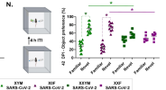
We used a spatial working memory task to measure short term hippocampal-dependent memory by quantifying spontaneous alternations in an adapted T-maze23,24 (Fig. 3A). As early as 7 dpi (Extended Data Fig. 3B) and continuing through 42 and 84 dpi (Fig. 3B), infected females had lower percentages of correct spontaneous alternation than either males or mock-infected females. Spatial working memory impairments in females were not due to differences in exploratory behavior, the number of arm entries (Extended Data Fig. 3C and Fig. 3C), or differences in the total distance travelled in the maze
The charts look quite convincing.

Other longer term behavioural impacts
Neither sex nor infection affected other behaviors analyzed, including exploratory behaviors, locomotor activity, anxiety-like behaviors (central tendency/rearing bouts in the open field), or depressive-like phenotypes (e.g., time spent immobile) in the tail-suspension test (Extended Data Fig. 3H-L). Infected females buried fewer marbles in the marble burying test as compared with either infected males or mock infected females, suggesting some alteration in compulsive-like behavior following infection, consistent with hamsters27 (Extended Data Fig.3M).
It's interesting that physical activity (exploratory behaviours, locomotor activity) didn't vary with infection (or sex). There's just that reduction in marble burying. It's not sounding as though there was a difference in most of the measures that might have captured fatigue.

There were persisting problems with loss of the sense of smell, but that didn't vary by sex.

They did the same studies with female mice infected with a more severe strain (they couldn't do the analyses in male mice because so many died).
Because neurological signs in females infected with ma10 we noted, we conducted a comprehensive behavioral and phenotypic SHIRPA assessment31. Ma10-infected females had reduced grip, motor, and coordination scores, increased latencies to descend a vertical pole, and numbers of grip slips during the pole climbing test compared to mu or mock-infected females, indicating that ma10 causes additional neurological manifestations (Extended Data Fig. 9H-I). Different SARS-CoV-2 variants result in shared female-biased PASC outcomes
That sounds as though the more severe strain did produce weakness and other motor symptoms, perhaps even fatigue.
 
There are aspects to the abstract that sound a bit too good to be true - such as finding things like neuroinflammation and dysregulated T cells that have not been clearly documented in humans, so much as speculated, as far as I know. But if the data are sound it makes sense and might be a foothold on to something important.

Confirming an X dosage rather than an absence of Y as the reason for female dominance would also be of major importance. A double dose of Xist and TLR-7 would make sense. And of course if gene dose is the culprit there is no need to do any GWAS or personal gene analysis - as far as I can see. The risk factor applies to all women equally. Polymohphisms may be irrelevant.
 
Last edited:
I suspect it doesn't actually matter too much if the 'PASC' they got isn't a terribly good model of the human illness. If the study shows that there is a sex difference in response to a virus that can explain why immune abnormalities persist in females despite males having worse acute disease it may be important.

It may also be that this is more a model of post-viral fatigue type illness than long term ME/CFS but if it provides a lead to understanding female predominance in ME/CFS that may not matter too much.

What I forget is how much post-viral fatigue is female dominant in humans though? Or even Long Covid? I suspect for Long Covid the figures are hard to be sure of because of the unhelpful diagnostic criteria being used.
 
Differences in host immune responses
(Just acknowledging the mice and the people who had to kill them to produce these results. Unlike some inane studies that involved killing animals, so far maybe this one warrants the sacrifice.)

Things seemed to be different by sex from very early on (2 dpi = 2 days post-infection)
In the lungs, 38/48 analytes were detectable, with 35/38 upregulated at 2 dpi in infected mice compared to sex-matched mock-infected controls (Table S1). Almost half (16/35) of the analytes that were increased at 2 dpi exhibited sex differences, with 15/16 analytes found in greater concentrations in infected females than males, including TNF-α, IL-6, IL-1β, CCL2, CCL3, and CCL4, which are involved in myeloid activation and recruitment (Fig. 4A and Table S1).
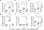
To assess sex differences in cellular responses to SARS-CoV-2, single cell suspensions were generated from spleens and myeloid subsets were evaluated during peak virus replication (Myeloid gating strategy 1, Extended Data Fig. 6A-F). Females had greater frequencies of inflammatory CCR2+ monocytes (Fig. 4B) and F4/80+ macrophages (Fig. 4C) at 2 dpi. While frequencies of CD86+ macrophages, CD206+ macrophages, neutrophils, and eosinophils were elevated at 2 dpi, sex differences were not observed (Extended Data Fig. 6B-F)

And at 7 dpi and 42 dpi
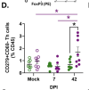
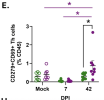
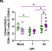
While frequencies of MHC II+macrophages (Fig. 4G) returned to back to baseline by 42 dpi, frequencies of monocytes (Fig.4D), CCR2+ monocytes (Fig. 4E), CD86+ macrophages (Fig. 4F), neutrophils (Fig. 4H), cDC1s (Fig. 4I), and pDCs (Extended Data Fig. 6H) were greater in infected females than males at 42 dpi. Despite a lack of persistent virus or viral RNA in extrapulmonary tissues, infected females had prolonged inflammatory phenotypes of innate immune cells as compared to males.
 
The shift in macrophage populations is interesting. This of course brings us right back to the lengthy discussion jnmaciuch and I had on which interferons from which cells are most likely to be involved in 'carrying over the feeling ill' from week to week, and year to year in ME/CFS in the absence of persistent virus.

I have been sceptical that there is a plausible way for shifts in macrophage populations to become established in some sort of carry-over cycle that perpetuates itself - i.e. a 'learnt' innate change. But I can see that if microarchitectural changes occurred in bone marrow, for instance, with reallocation of niches in response to TLR7 signalling, those niches might get fixed for long periods so that the monocyte output was skewed for months or even years.

I am reminded of the mystery of the B cell depletion period after rituximab which can last anything from 6 months to five years despite the drug having largely disappeared by about two months. In a way it is a variant of the writing on the wall story - a change in parking restriction notices that only get changed every five years.
 
Another thought is that although a role for TLR7 in female preponderance in autoimmune diseases could easily involve interferon pathways that need not mean that a role for TLR7 in ME/CFS makes it autoimmune. The same basic difference in interferon handling may generate disease through two quite different routes. Lupus might use both routes!
 
No difference in viral quantities
They didn't find differences in the quantity of virus in tissues.
This would seem to be more evidence against viral persistance driving symptoms then. If only we could get the US LC research lot to listen...
There are aspects to the abstract that sound a bit too good to be true - such as finding things like neuroinflammation and dysregulated T cells that have not been clearly documented in humans, so much as speculated, as far as I know.
But were these found during autopsies of the mice and/or in tissues that can't safely be tested in live humans?
 
But were these found during autopsies of the mice and/or in tissues that can't safely be tested in live humans?

The brain bit might be. I am not sure it matters. What worries me is that the study seems to have found all the things we might predict it would find based on popular ideas. I haven't read the whole paper yet as I am supposed to be doing a ForwardME meeting this morning but I get the impression from Hutan's scan that they have done this pretty thoroughly.
 
Back
Top Bottom