IFNγ causes mitochondrial dysfunction and oxidative stress in myositis, 2024, Abad et al.

jnmaciuch

Senior Member (Voting Rights)

IFNγ causes mitochondrial dysfunction and oxidative stress in myositis​

Catalina Abad, Iago Pinal-Fernandez, Clement Guillou, Gwladys Bourdenet, Laurent Drouot, Pascal Cosette, Margherita Giannini, Lea Debrut, Laetitia Jean, Sophie Bernard, Damien Genty, Rachid Zoubairi, Isabelle Remy-Jouet, Bernard Geny, Christian Boitard, Andrew Mammen, Alain Meyer & Olivier Boyer

Abstract

[Paragraph breaks added]

Idiopathic inflammatory myopathies (IIMs) are severe autoimmune diseases with poorly understood pathogenesis and unmet medical needs. Here, we examine the role of interferon γ (IFNγ) using NOD female mice deficient in the inducible T cell co-stimulator (Icos), which have previously been shown to develop spontaneous IFNγ-driven myositis mimicking human disease.

Using muscle proteomic and spatial transcriptomic analyses we reveal profound myofiber metabolic dysregulation in these mice. In addition, we report muscle mitochondrial abnormalities and oxidative stress in diseased mice. Supporting a pathogenic role for oxidative stress, treatment with a reactive oxygen species (ROS) buffer compound alleviated myositis, preserved muscle mitochondrial ultrastructure and respiration, and reduced inflammation. Mitochondrial anomalies and oxidative stress were diminished following anti-IFNγ treatment.

Further transcriptomic analysis in IIMs patients and human myoblast in vitro studies supported the link between IFNγ and mitochondrial dysfunction observed in mice. These results suggest that mitochondrial dysfunction, ROS and inflammation are interconnected in a self-maintenance loop, opening perspectives for mitochondria therapy and/or ROS targeting drugs in myositis.

Link (open access)
 
will come back to do an "explain like I'm brain foggy" write-up and discuss parts of the paper later. Ended up at this paper trying to follow a thread after seeing that "flu-like fatigue" is commonly reported in myositis


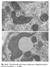
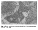
I was also trying to find images of mitochondrial morphology under various conditions to see how they compared to Behan et al. 1991
 
Explain like I'm brain-foggy:
Myositis is an autoimmune condition of the muscle. A mouse model of type I diabetes missing a specific T cell gene (Icos) is known to spontaneously develop myositis, and previous studies have confirmed that myositis in these mice requires interferon gamma signaling.

Transcriptomics showed that both interferon gamma and beta reached high levels (detected by mRNA) only in the disease state, which occurs at around 25 weeks of age in these mice. Note that this study did not seem to distinguish between the downstream effects of interferon beta vs. interferon gamma at any step, so we can't directly attribute effects to one or the other.

The transcriptomic analysis also showed that many mitochondrial and metabolism-related genes were differentially expressed in the Icos knockout. They also found differences in metabolic capacity correlating with grip strength and gait, substantial differences in mitochondrial morphology, and elevated ROS production.

Finally, the authors showed that preventive treatment with JAK-STAT inhibitors (blocking the interferon response) prevented the mitochondrial abnormalities in these mice (as well as overall disease, which was expected). They also showed that they could induce those mitochondrial abnormalities in cultured muscle cells by adding interferon gamma. Preventitive anti-ROS treatment somewhat ameliorated disease severity and mitchondrial abnormalities, but not to the same extent as the anti-interferon therapies.

Because of the peculiarities of this mouse model, I think comparisons to human myositis should still be taken with a grain of salt. However, this study is still useful because it proves that abnormal interferon signaling can directly cause mitochondrial abnormalities, and that these mitochondrial abnormalities seem to further drive the interferon response.
 
Last edited:
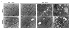
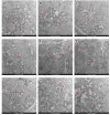
Taking a deeper look at that mitochondrial morphology:
1751241741555.png
The three columns on the right are from the myositis model. To my amateur eye, you can see what looks to be vacuolization (the white spots), pleomorphy aka substantial irregularity in size (particularly in that bottom right example), and cristae disorganization.

1751241899416.png
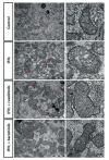
The second row shows mitochondria from muscle cell culture with interferon gamma added. You see that vacuolization and what sorta looks like "compartmentalization" in the inner matrix. The bottom two rows show mitochondria in the JAK-STAT inhibitor conditions, demonstrating that blocking the interferon response makes the mitochondria resemble control.

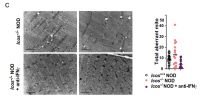
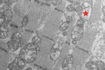
I'm less convinced by this supplemental figure, which is showing the same view as the first figure in this post, now testing the effect of JAK-STAT inhibitors.
1751242136831.png
You can see the bars on the right showing increased number of "abnormal" mitochondrial in the Icos knockout without treatment, but the variability is huge (and they didn't report a p-value). I would've liked to see quantification for the other two imaging figures so I can be confident that it's a consistent trend beyond these representative images.


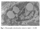
And as comparison, the images from Behan et al. 1991:
1751242321009.png1751242372417.png1751242393986.png

Lots of things can cause changes in mitochondrial morphology so this isn't proof that interferon is causative in the post-viral ME cases from Behan et al. I just thought it was interesting to see some of the overlap in morphological abnormalities
 
Those Behan images look compelling as far as a story of vacuolisation goes, and I'd love there to be a difference as obvious as that, but I can't recall seeing that finding reported elsewhere?

Surely if those vacuoles were a common feature of ME/CFS muscle cell mitochondria, there would have been more reports of them?
 
I am working on my phone today but those images don't look that similar to me. Interpretation of histology is crucially dependent on what you see nearby an "abnormality" or even more importantly what is no there, like a T cell for instance.

Human autoimmune myositis is almost certainly mediated by antibody rather than T cells but there may be interferon generated secondarily. I agree with @jnmaciuch 's caveats. But it is interesting to see what IFNs can do.
 
Similar in cardiac muscle cells. SARS-CoV-2 damages cardiomyocyte mitochondria and implicates long COVID-associated cardiovascular manifestations (2025). Just five patients (with myocarditis) —

Electron microscopy further detailed local myofibrillar fibrosis, fibroblast proliferation, muscle bundle edema, loss of myofibrillar integrity, presence of lipofuscin granules in cardiomyocytes, and significant mitochondrial vacuolation (refers loss of cristae structure and formation of vacuoles or vacuole-like mitochondria,

We have observed an interesting phenomenon in the mitochondria, the swollen and vacuolated mitochondria, distorted and broken cristae, all indicated severe damage to this important cellular organelle. This damage is highly likely caused by the SARS-CoV2 Infection, as we have found an almost identical mitochondria disorganization in the mice that were infected with SARS-CoV-2.

Screenshot 2025-06-30 at 10.16.38 PM copy.jpgScreenshot 2025-06-30 at 10.16.59 PM copy.jpg
 
However, this study is still useful because it proves that abnormal interferon signaling can directly cause mitochondrial abnormalities, and that these mitochondrial abnormalities seem to further drive the interferon response.

See also Core mitochondrial genes are down-regulated during SARS-CoV-2 infection of rodent and human hosts (2023) —

At high viral load, the most notable changes in mitochondrial gene expression were the strong suppression of ALAS2, one of the 5'-aminolevulinate synthetases involved in heme synthesis; strong induction of the complex IV assembly factor, SCO2; and up-regulation of the mitochondrial genes thought to be important in the mitochondrial induction of the innate immune response.

These virally induced mitochondrial innate immune regulatory genes included PNPT1 (polyribonucleotide nucleotidyltransferase 1), which may release mitochondrial double-stranded RNA to engage melanoma differentiation-associated protein 5 (IFIH1) and trigger the type I interferon response; CMPK2 (cytosine monophosphate kinase 2), which is the rate-limiting step for the induction of mtDNA replication creating oxidized mtDNA (Ox-mtDNA), which is released from mitochondria and binds to the inflammasome; ACOD1 (aconitate decarboxylase 1 or IRG1), which regulates itaconate production and modulates inflammatory myeloid cells; and IFI27 (interferon alpha inducible protein 27), which activates interferon expression and is a diagnostic blood marker for SARS-CoV-2 infection.

Hence, SARS-CoV-2 not only inhibited OXPHOS but also activated mitochondrial innate immunity signaling genes (CMPK2, PNPT1, IFI27, and IRG1)
 
Back
Top Bottom