Haptoglobin phenotypes and structural variants associate with post-exertional malaise and cognitive dysfunction in [ME], 2025, Moezzi, Moreau et al

Nightsong

Senior Member (Voting Rights)
Abstract:

Background​

Myalgic encephalomyelitis (ME) is a chronic, multisystem illness characterized by post-exertional malaise (PEM) and cognitive dysfunction, yet the molecular mechanisms driving these hallmark symptoms remain unclear. This study investigated haptoglobin (Hp) as a potential biomarker of PEM severity and cognitive impairment in ME, with a focus on Hp phenotypes and structural proteoforms.

Methods​

A longitudinal case–control study was conducted in 140 ME patients and 44 matched sedentary healthy controls. In the discovery phase, global plasma proteomic profiling was performed in 61 ME patients and 20 controls before and after a standardized, non-invasive stress protocol in order to induce PEM. Associations between Hp levels, phenotype, and cognitive performance were assessed. In the validation phase, plasma Hp concentrations and proteoform composition were analyzed in an independent cohort of 89 ME patients and 24 controls using high-performance liquid chromatography (HPLC).

Results​

ME patients demonstrated a significant reduction in Hp levels following post-exertional stress. Lower baseline Hp concentrations were associated with impaired cognitive performance. Hp phenotypes were differentially associated with symptom burden, with the Hp2-1 phenotype enriched in ME and linked to greater PEM severity and cognitive deficits compared to Hp1-1 and Hp2-2. HPLC analysis revealed altered Hp proteoform profiles in the Hp2-1 subgroup, including increased high-mass tetrameric and pentameric forms and shorter retention times indicative of structural changes. In contrast, the Hp1-1 phenotype was associated with milder symptoms and greater cognitive resilience.

Conclusions​

These findings suggest that Hp phenotype and proteoform structure modulate the physiological response to post-exertion in ME, offering insight into the molecular basis of PEM and its clinical heterogeneity. Hp may serve as a translational biomarker for patient stratification and a potential therapeutic target to mitigate oxidative stress and cognitive dysfunction in ME.

 
Proteomic analysis revealed that Hp levels were significantly higher at baseline compared to 90 min post-stress in the ME group (1.84-fold change, p = 0.007). This stress-induced decrease in Hp level was specific to ME patients and was not observed in healthy controls, in whom Hp levels remained undetectable at both time points.
The pronounced post-exertional reduction in Hp, a key scavenger of free hemoglobin, in ME patients suggests a diminished capacity to buffer hemolysis-associated oxidative and inflammatory stress.

Supporting this, the non-invasive cerebral monitoring using MASIMO's NIRS revealed a significant post-PEM increase in cerebral total hemoglobin in ME patients, a pattern absent in healthy controls (Supplementary Figure S4).

Importantly, our cohort was composed primarily of severely affected, housebound individuals, patients who are rarely included in clinical studies due to the demands of protocols such as cardiopulmonary exercise testing (CPET), which typically excludes this population.

The standardized 90-min provocation maneuver used here was carefully tailored to this severity level and, based on extensive clinical experience, is sufficient to trigger PEM in this group.
 
Last edited:
Myalgic encephalomyelitis (ME) is a chronic, disabling disease characterized by profound fatigue, unrefreshing sleep, and cognitive impairment. The hallmark symptom, post-exertional malaise (PEM), involves exacerbation of symptoms following minimal exertion (physical or mental) and lacks a clear mechanistic explanation [1, 2].
PEM severity was further evaluated using a modified version of the DePaul Post-Exertional Malaise Questionnaire (DPEMQ), which was administered by a clinical research nurse five days after the stress test [18]. This version focused on the frequency and severity of symptoms during the immediate post-exercise period rather than over the previous six months. Thirteen items contributed to the total DPEMQ score, with higher scores indicating greater PEM severity.
I think 18 is this:

I don’t fully understand how they measured PEM or what they mean by it. I wish they spent more effort on explaining their understanding of PEM and showing a timeline.

They say the controls were sedentary, but I can’t find any data on activity levels. Can we exclude the possibility that the observed differences are due to very inactive muscles being stimulated compared to somewhat active muscles being stimulated?
 
Can we exclude the possibility that the observed differences are due to very inactive muscles being stimulated compared to somewhat active muscles being stimulated?

I wondered that.

It would have been good to include some moderately ill people too; in terms of how much they use the arm muscles being stimulated, they'd probably match sedentary controls quite well. But if a project can only include one cohort due to funding limitations, it is important to prioritise severely ill people.
In principle I agree that prioritising the severe is important, but only if it doesn’t reduce the value of the information you get from the study.

What they were trying to figure out is if the body of pwME/CFS reacts abnormally to external stimuli of the muscles in the arm. If you are unable to control the large confounder of baseline activity for that muscles group when studying the severe, wouldn’t it be better to study moderate or even mild patients first, and then move on to the severe to see if the findings replicate there?

Unless you’ve got a reason to think that the severe are somehow different from the moderate/mild, in other ways than just being more sick with the same thing?
 
If you are unable to control the large confounder of baseline activity for that muscles group when studying the severe, wouldn’t it be better to study moderate or even mild patients first, and then move on to the severe to see if the findings replicate there?
On the other hand, we're talking about a disease where proteomics and exercise tests have already been tried several times in less severe people. If there actually are differentially expressed proteins, we expect them to probably have tiny effect sizes, and when you're correcting for a hundred tests, you might miss them.

So it might be better to compare to people who are severe, to get as large of effect sizes as possible, so they aren't missed. Then try to replicate just the proteins of interest in groups that better match.
That’s a good point.

My main worry is that you’ll just end up finding noise all the time due confounders and chasing red herrings. Maybe a three group setup would be better - HC, severe and moderate/mild, or HC, severe and another disease?
 
That’s a good point.

My main worry is that you’ll just end up finding noise all the time due confounders and chasing red herrings. Maybe a three group setup would be better - HC, severe and moderate/mild, or HC, severe and another disease?
I'm sure that's been done a few times. And quite a few of those severe ended up halfway between health controls and mild/moderate, sometimes with generous explanations of how this makes sense. Which makes me wonder if it's a case of more red herring is.
 
September 16, 2025

OMF’s Collaborative Center at Montreal, in collaboration with our Collaborative Center at Uppsala and the Computational Research Center for Complex Diseases, has released a new paper: Haptoglobin phenotypes and structural variants associate with post-exertional malaise and cognitive dysfunction in myalgic encephalomyelitis.

From the Desk of Dr. Alain Moreau, Director of the ME/CFS Collaborative Research Center at Montreal:

Dr. Alain Moreau, wearing a blue suit and glasses, smiles as he points to a scientific poster in a research lab.
What is this study about?


This study investigated a blood protein called haptoglobin (Hp), which normally protects the body from damage when red blood cells break down. OMF researchers from Montreal, Uppsala, and the OMF computation team looked at how Hp levels and genetic variants behave in people with ME/CFS, especially during post-exertional malaise (PEM)—the worsening of symptoms after even minor activity.

Why does it matter?

They found that after a mild stress challenge, people with ME/CFS showed a sharp drop in Hp levels, while healthy controls did not. Low Hp was linked to worse PEM symptoms and poorer cognitive performance (“brain fog”). Importantly, the study also revealed that certain genetic forms of Hp influence how severe PEM becomes: patients with the Hp2-1 genotype had stronger PEM and more cognitive problems, while those with Hp1-1 were more resilient. This shows that genetics directly contribute to vulnerability in ME/CFS.

What is the potential impact for people with ME/CFS?

This is the first strong evidence of a genetic contribution to PEM in ME/CFS. Hp could serve as a biomarker to better diagnose ME/CFS, identify patient subgroups, and guide clinical management. In the future, therapies that restore or replace Hp function (e.g., intravenous infusion of recombinant Hp-1-1 protein) might help protect genetically at-risk patients from post-exertional crashes and cognitive decline, offering a new pathway toward targeted treatment.

Read the full publication in the Journal of Translational Medicine.
 
Among the proteins altered in response to stress, Hp emerged as the most significantly dysregulated in ME patients (Table 2, Fig. 2 and Supplementary Figure S6). Proteomic analysis revealed that Hp levels were significantly higher at baseline compared to 90 min post-stress in the ME group (1.84-fold change, p = 0.007). This stress-induced decrease in Hp level was specific to ME patients and was not observed in healthy controls, in whom Hp levels remained undetectable at both time points. This distinct post-stress pattern positions Hp as a potential disease-specific marker of PEM. The pronounced post-exertional reduction in Hp, a key scavenger of free hemoglobin, in ME patients suggests a diminished capacity to buffer hemolysis-associated oxidative and inflammatory stress.
The paper says that Hp levels were undetectable before and after the stressor in the healthy controls. But for the ME group, HP was present at baseline, but decreased after the stressor.

To me, the situation as described there for the ME/CFS group is normal. It is not normal to have no Hp.
AI said:
Normal haptoglobin (Hp) levels in adults generally range from about 40 to 200 mg/dL, but this can vary slightly depending on the laboratory and individual factors like age and gender.

Reading on, there is an explanation.
The identification of Hp as a top candidate biomarker warrants careful consideration in light of the technical protocol employed. The plasma samples used in the global proteomic assay were processed using a MAR-14 immunodepletion column, which targets and removes high-abundance plasma proteins, including acute-phase reactants such as Hp. In theory, this step should have substantially reduced or eliminated detectable Hp levels across all samples. The fact that Hp remained quantifiable among the ME group, and showed consistent, biologically plausible differences post-stress, raises important questions about the molecular form and binding context of the Hp detected.

So, actually the processing was supposed to remove "high-abundance plasma proteins" including Hp. So, the processing explains why there was no Hp in the healthy controls. It doesn't explain why Hp was found in the ME/CFS group. It seems possible that there was a lab error. They are suggesting that the difference might be related to some difference in ME/CFS Hp.

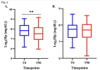
To validate and extend the discovery phase results obtained using MAR-14-depleted plasma proteomics, a high-sensitivity method for direct Hp quantification in total plasma (without prior depletion) was applied to the same cohort. This orthogonal approach confirmed a significant post-stress reduction in Hp levels in ME patients, with the medium effect size (Fig. 4). In contrast, no significant change in Hp levels was found in HC group, confirming the disease specificity of the stress-induced Hp depletion (Fig. 5). This replication strengthens the evidence that Hp is a stress-responsive protein in ME and may be a biomarker of PEM.
So, there they are saying that they used another method, this time directly quantifying Hp levels in the plasma. Figure 5 gives those actual levels.
Screenshot 2025-09-17 at 1.15.45 pm.png

Chart A is for the ME/CFS group; Chart B is for the control group. Note the different y axis scale. Really, there's nothing much to the claimed differences between the ME/CFS and control groups, and even from before and after the stressor. Maybe the stressor just isn't enough to make a difference?

It is normal for levels to decrease a bit after exercise, so that very small change found in the ME/CFS group is not remarkable.

Perhaps there is something about ME/CFS Hp that resulted in it still being present after that first processing approach that should have removed it. It's worth looking into more. But, it could easily just be some sort of an error, some difference in the sample processing.
 
Back
Top Bottom