Review Fluid transport in the brain, 2022, Rasmussen, Mestre, Nedergaard

SNT Gatchaman

Senior Member (Voting Rights)
Staff member
Fluid transport in the brain
Martin Kaag Rasmussen; Humberto Mestre; Maiken Nedergaard

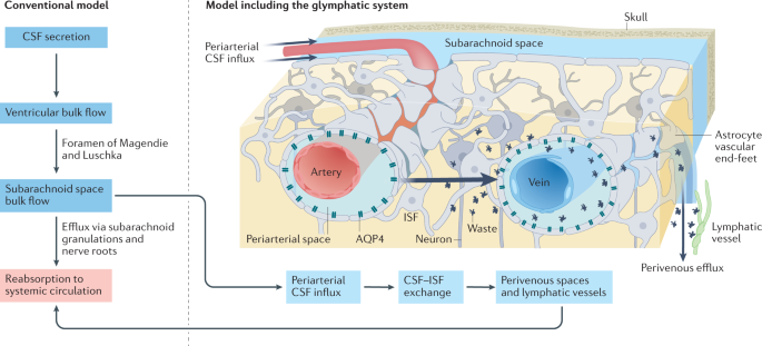
The brain harbors a unique ability to, figuratively speaking, shift its gears. During wakefulness, the brain is geared fully toward processing information and behaving, while homeostatic functions predominate during sleep. The blood-brain barrier establishes a stable environment that is optimal for neuronal function, yet the barrier imposes a physiological problem; transcapillary filtration that forms extracellular fluid in other organs is reduced to a minimum in brain. Consequently, the brain depends on a special fluid [the cerebrospinal fluid (CSF)] that is flushed into brain along the unique perivascular spaces created by astrocytic vascular endfeet.

We describe this pathway, coined the term glymphatic system, based on its dependency on astrocytic vascular endfeet and their adluminal expression of aquaporin-4 water channels facing toward CSF-filled perivascular spaces. Glymphatic clearance of potentially harmful metabolic or protein waste products, such as amyloid-β, is primarily active during sleep, when its physiological drivers, the cardiac cycle, respiration, and slow vasomotion, together efficiently propel CSF inflow along periarterial spaces. The brain’s extracellular space contains an abundance of proteoglycans and hyaluronan, which provide a low-resistance hydraulic conduit that rapidly can expand and shrink during the sleep-wake cycle.

We describe this unique fluid system of the brain, which meets the brain’s requisites to maintain homeostasis similar to peripheral organs, considering the blood-brain-barrier and the paths for formation and egress of the CSF.

Web | DOI | PDF | Physiological Reviews | Open Access
 
The blood-brain barrier establishes a stable environment that is optimal for neuronal function, yet the barrier imposes a physiological problem; transcapillary filtration that forms extracellular fluid in other organs is reduced to a minimum in brain. Consequently, the brain depends on a special fluid [the cerebrospinal fluid (CSF)] that is flushed into brain along the unique perivascular spaces created by astrocytic vascular endfeet.

This, surely, is utter nonsense. All fluid in the brain parenchyman has to come from transcapillary filtration. The fluid that comes from transcapillary filtration in choroid plexus merely passes through the ventricular system, it does not wash through deep brain parenchyma.
 
Last edited:
I am still very puzzled by this. I am prepared to believe that they are describing a real fluid flux system but I cannot see how it works.

My first query is about the claim tht water flow is reduced to a minimum because of the blood brain barrier. (It might be minimal, but for other reasons such as positive CSF pressure.) Water molecules are smaller than oxygen and carbon dioxide molecules, which have to zip across the BBB continually. Is the BBB really impervious to water?

The other puzzle is the route for the glymphatic. I tried reading the review but got bogged down because it did not give a simple explanation at the beginning. Brain tissue is fed by blood vessels and drained by blood vessels. In all other tissues these run together until they become large arteries and veins, so at the level of fluid flux, i.e. capillaries and venules, there is only one perivascular space around both the in and the out vessels. So if glymphatics run in this space there is no through route, only the same route in as out. Pumping fluid along a perivascular space with a blind end would be an extremely inefficient method of 'flushing' because the fluid would have to come back out the same way. Rapid shunting phases would just push to fluid back and forth a bit without achieving much. This cannot be what is proposed but I have yet to understand what IS proposed.
 
Water molecules are smaller than oxygen and carbon dioxide molecules, which have to zip across the BBB continually. Is the BBB really impervious to water?
It’s primarily through aquaporins if I’m remembering correctly. Passive diffusion is also possible depending on various factors contributing to permeability but accounts for a much lower percentage of total water influx

[Edit: cross-posted with @Kitty ]
 
Is it possible the movement is only one way?

It's just that the diagram suggests CSF goes in via arterial spaces and exits via venous ones—but interstitial fluid is only shown on the out route.

(The diagram's got nothing to do with this study tho.)
 
It's just that the diagram suggests CSF goes in via arterial spaces and exits via venous ones—but interstitial fluid is only shown on the out route.

Interstitial fluid only on the out route would be the old theory - coming in through the blood vessels. If it comes in alongside arteries how would the pump work - since the arteries come in from underneath in the circle of Willis, not through the CSF ventricles? And it is hard to see how pushing fluid in along arteries helps push it out along veins? In between there are capillaries with no perivascular space.
 
To be honest I only looked it up because wanted to know what astrocytic vascular endfeet were. The pictures didn't disappoint, though I still think the name is a tautology.
 
What are aquaporins? And what is primarily through them? And from where to where? I want to understand the anatomical routes.
Aquaporins are channels in cell membranes that can open to allow selective passage to water and other solutes. Present on nearly all cells, but especially on endothelial/epithelial cells of blood vessels and astrocytes.

What's being described is: blood vessels project into the brain parenchyma. In other tissues, solutes can exit the blood vessel directly into the parenchymal interstitial space. But around each blood vessel in the brain, there is an additional sheath, made of up the endfeet of astrocytes.

The space between the blood vessel wall and the astrocytic endfeet is the "perivascular" space. The fluid in this perivascular space is what is referred to as the "lymphatic fluid" in the brain.

Water and other solutes from this lympathic fluid enter the actual body of the astrocytes via aquaporins on these endfeet. Water and solutes can then exit the astrocyte into the parenchymal space via aquaporins on other parts of the astrocyte. The astrocytic body effectively serves as a customs area--intracellular regulation of aquaporins/other channel proteins can regulate which solutes and how much pass through either end.
 
Last edited:
Brain CFS flow.webp
Cerebrospinal fluid (CSF) flow according to the conventional view (left) and including the glymphatic model (right). According to the conventional model, CSF is produced in the ventricles and flows to the subarachnoid space to be directly reabsorbed into the bloodstream. In the updated glymphatic model, part of CSF flows into the brain along the perivascular spaces of penetrating arteries. In a high glymphatic flow state (for example, natural sleep), CSF enters brain tissue supported by aquaporin 4 (AQP4) water channels. CSF mixes with interstitial fluid (ISF) and drains from the brain along perivenous spaces. The elongated and elliptical shape of the periarterial spaces and the eccentric localization of arteries within the periarterial spaces reduce resistance to fluid flow. Although peripheral arteries and veins generally occur side by side, along with nerves, blood vessels of the central nervous system (CNS) are not confined together in fascia but located separately and follow distinct trajectories. This unique organization of vasculature in the CNS has been hypothesized to generate a pressure gradient for fluid flow from the periarterial to the perivenous spaces through the interstitium.

This is the reference for how the fluid flows across the ISF:
 
So then the argument re: the glympathic system and waste clearance is that some (but likely not all) waste products don’t pass back through the endothelial right junction of the blood vessel endothelium into the circulation, they just pass through astrocytes into the “brain lymph” and get cleared out from there?

[Edit: I guess that’s where the discussion of dorsal dura mater lymphatic vessels comes in to answer the question of “clears out to where?” Seems like an incompletely answered question]
 
Last edited:
So then the argument re: the glympathic system and waste clearance is that some (but likely not all) waste products don’t pass back through the endothelial right junction of the blood vessel endothelium into the circulation, they just pass through astrocytes into the lymph and get cleared out from there?

[Edit: I guess that’s where the discussion of dorsal dura mater lymphatic vessels comes in to answer the question of “clears out to where?” Seems like an incompletely answered question]

From what I can understand it seems like after moving from the periarterial space to the perivenous space it flows back into the subarachnoid space. From here CFS is being reabsorbed into the superior sagittal sinus through arachnoid granulations which takes some "waste" with it. Lymphatic vessels also directly remove the CFS from the perivenous space removing more of the fluid.

The fluid in the subarachnoid space would contain waste from the perivenous efflux side. So the fluid that the astrocytes move across would already have some waste in it. Fresh CFS is constantly being produced and old CFS removed from the subarachnoid space. Given that waste comes from the ISF, then it would seem that the concentration of waste in the ISF will be higher than the waste in the CFS (as you are constantly adding some clean fluid into the system). So when the fluid goes from the periarterial to the perivenous side the concentration of waste increases. The net outflow of waste would just depend on how much fresh CFS was entered into the system and how much waste is in the CFS that is removed from the system.

Maybe there are some ways that the outflow from the perivenous space can be more efficiently funneled to lymphatics or into the venous system but I can't find much on that. Another thought might be that astrocytes can "block" some of the waste from entering the ISF when traveling from the periarterial to the perivenous side creating much more of a "waste" gradient.
 
The other puzzle is the route for the glymphatic. I tried reading the review but got bogged down because it did not give a simple explanation at the beginning. Brain tissue is fed by blood vessels and drained by blood vessels. In all other tissues these run together until they become large arteries and veins, so at the level of fluid flux, i.e. capillaries and venules, there is only one perivascular space around both the in and the out vessels. So if glymphatics run in this space there is no through route, only the same route in as out. Pumping fluid along a perivascular space with a blind end would be an extremely inefficient method of 'flushing' because the fluid would have to come back out the same way. Rapid shunting phases would just push to fluid back and forth a bit without achieving much. This cannot be what is proposed but I have yet to understand what IS proposed.

After spending some time looking through the paper there is still a lot I don't understand but I thought I'd send some of the parts that seemed important. I also don't know if what I said in the second paragraph above is even somewhat accurate.

The first thing the paper seems to suggest is that the CFS entering into the brain enters alongside the arterial system

Cerebrospinal fluid(CSF) enters the brain along the arterial vascular system of the brain that consists primarily of the 3 large cerebral arteries: the anterior cerebral artery(ACA), the middle cerebral artery (MCA), and the posterior cerebral artery (PCA)
Brain Fluid transport.png

In terms of the movement of fluid along the glymphatics:

The glymphatic system conceptualizes brain fluid transport into three segmental fluid transport pathways: 1) periarterial CSF influx, 2) AQP4-supported influx and dispersion of CSF in the extracellular space, and 3) perivenous efflux. Meningeal lymphatic vessels surround the large venous sinuses and export, at least in part, perivenous fluid, thereby adding a fourth segment to brain fluid transport.

The CSF that enters the periarterial spaces is pumped forward in an anterograde manner along the major cerebral arteries and from there it enters the brain through the perivascular spaces of the thousands of penetrating arteries that perforate the brain tissue ... The perivascular spaces are open and provide little resistance for CSF influx driven by arterial pulsatility

But later they say this:

There are two main models of perivascular transport: those consisting of anterograde flow into the brain within the perivascular space and those evaluating retrograde flow out of the brain within basement membranes. In both cases, the general consensus is that arterial pulsations alone are not sufficient to drive net flow in any direction, thus contradicting experimental results that show bulk flow is in the direction of blood flow. Studies have proposed that an as yet unknown pressure gradient or the presence of a valve-like mechanism could explain these opposing results.

For the movement of fluids within the extracellular space
AQP4 expression plays a critical role in regulating intra axial fluid flow. Global AQP4 knockout mice have reduced influx of CSF tracers compared with wild-type animals. Likewise, CSF tracers injected into the brain parenchyma of Aqp4-/- mice have slower clearance than in wild-type mice. Adequate polarization to the end feet seems to be critical for facilitating fluid transport, since knockout animals that have preserved AQP4 expression but lack polarization have reduced tracer transport.
The flow of fluid through the tortuous brain extracellular space is probably driven by a combination of diffusion and convection.
The observation that CSF enters along arterial perivascular space and subsequently shows up at the venous perivascular space indicates that CSF influx might be a direct driver for extracellular fluid efflux. There are several possible scenarios for such a mechanism: 1) the flow of CSF into arterial perivascular space continues down along the capillary, either through an existing perivascular space or along channels created by basement membranes, eventually connecting with the venous side perivascular space; 2) the entirety of the flow continues into the extracellular space and re-enters the venous perivascular space at some downstream location
If flow is to continue along the perivascular space, there must be a pressure gradient from the vicinity of the artery toward the venous side. Such a pressure gradient along the arterial/venous perivascular space has not yet been measured, but there are several models for how it might occur. The best developed theory is that CSF flow is driven by perivascular (peristaltic) pumping caused by the arterial wall pulsation of the cardiac cycle
 
What's being described is: blood vessels project into the brain parenchyma. In other tissues, solutes can exit the blood vessel directly into the parenchymal interstitial space. But around each blood vessel in the brain, there is an additional sheath, made of up the endfeet of astrocytes.

The space between the blood vessel wall and the astrocytic endfeet is the "perivascular" space. The fluid in this perivascular space is what is referred to as the "lymphatic fluid" in the brain.

Yes, I get that much, but it doesn't address the in-out anatomy issue
Aquaporins are channels in cell membranes that can open to allow selective passage to water and other solutes. Present on nearly all cells, but especially on endothelial/epithelial cells of blood vessels and astrocytes.

I suspected as much.
Water and other solutes from this lympathic fluid enter the actual body of the astrocytes via aquaporins on these endfeet. Water and solutes can then exit the astrocyte into the parenchymal space via aquaporins on other parts of the astrocyte. The astrocytic body effectively serves as a customs area--intracellular regulation of aquaporins/other channel proteins can regulate which solutes and how much pass through either end.

OK, so there is an outer barrier as well as the vascular endothelium barrier, but this outer barrier has holes in. How does this help the flux question? If the water is not coming out of blood vessels and is coming via glymphatics and then into parenchyma what makes it come back out of parenchyma into the same glymphatics and why doesn't it ust go back where it came?
.
 
Studies have proposed that an as yet unknown pressure gradient or the presence of a valve-like mechanism could explain these opposing results.

This is what is missing. I see what they are trying to propose now, but it is crucially dependent on a valve mechanism, which I didn't see mentioned at the start, or some clever pressure gradiet from arterial pulsation, which they seem to discount.

Interesting to know what aquaporins are but I cannot see how they contribute anything of interest. And if the water can only get through tiny holes there would have to be either a valve system in the astrocytes with active transport or a decent hydrostatic gradient - which seems to be missing at that scale.

What worries me is that all the stuff about electrical brain activity pumping water through makes no real sense unless you have a valve system. If you have a valve system then anything that tends to squeeze and unsqueeze will drive fluid through - that's easy and you don't need to postulate anything terribly clever about the perivascular space at all - except valves.

The whole thing looks pretty inefficient to me and I continue to wonder whether they are just studying an epiphenomenon - like an artery in the crook of the elbow wiggling from side to side with pulse for no particular purpose. I can imagine that any jiggling of brain parenchyma might send water through if there were valves but is it really iportant. Metabolic waste products will cross back into venules happily. Degraded proteins like bundles of misfolded amyloid could easily be pinocytosed and metabolised by microglia. (The theory seems to be that you need to wash them out to get them to macrophages for eating in lymph node, so why not just let microglia do it?)

And what I don't get is why sleep should be important since arterial pulsation, valves and 'waves' of electrical activity are going on nicely during the day. Why would you need to remove amyloid at night?
 
I am still sceptical about the idea that this is all because the BBB stops water coming out of blood vessels. A much simpler explanation is that the oncotc gradient is higher because of the barrier to protein flux and the CSF pressure is positive, whereas interstitial pressure in other tissues is subatmospheric because there is a sponge and valve effect there. Starling forces across brain vessels may well be zero - whereas for other tissues they never were and the student-teaching version of Starling was wrong, as we worked out in the 1970s.
 
OK, so there is an outer barrier as well as the vascular endothelium barrier, but this outer barrier has holes in. How does this help the flux question? If the water is not coming out of blood vessels and is coming via glymphatics and then into parenchyma what makes it come back out of parenchyma into the same glymphatics and why doesn't it ust go back where it came?
Flow through aquaporins is usually bidirectional and dictated by osmotic gradient. Aquaporins on astrocytes make up a large proportion of the membrane surface area so it seems that it really is a matter of free flow for water itself, but not other solutes that don’t have a comparable selective channel. Aquaporins would also be present on the brain vessel endothelium—I’m guessing the comment about water being unable to cross the BBB more specifically refers to a lack of passage through gaps in endothelial layers. Water is still crossing it’s just controlled through the cell bodies

It’s also known that astrocytes can swell and control their own volume dynamically, so perhaps that is part of the picture. The ones positioned at various parts of the inbound/outbound routes may have ways of adjusting their own internal osmolarity. Perhaps this is enough to address your questions of flux, perhaps not

Degraded proteins like bundles of misfolded amyloid could easily be pinocytosed and metabolised by microglia. (The theory seems to be that you need to wash them out to get them to macrophages for eating in lymph node, so why not just let microglia do it?)
As far as I know, amyloid clearance has been observed to occur through multiple methods—extracellular cleavage from glial cell-derived proteins, pinocytosis and internal degradation via lysomomes or proteasome, chaperoning of AB fragments into the circulation, etc. So I don’t think the argument is that glympathic clearance is the only or even the most important method. It’s just that various methods operate at their own capacities, and there may be some instances where insufficient clearance via one route cannot be adequately compensated by others. Even if pinocytosis by microglia could technically handle all clearance, amyloid uptake induces transcriptional and metabolic changes that may make microglia less capable of carrying out other vital functions, so it may have been evolutionarily beneficial to distribute the labor across other systems
 
Flow through aquaporins is usually bidirectional and dictated by osmotic gradient. Aquaporins on astrocytes make up a large proportion of the membrane surface area so it seems that it really is a matter of free flow for water itself, but not other solutes that don’t have a comparable selective channel.

But that doesn't sound much use. The water is supposed to be taking the bad solutes out with it. And I don't see any particular need to stop solutes going with the water on the upstream side. And since there appear to be no osmotic or hydrostatic gradients here much it is a bit hard to know what the point of special holes is.

My impression so far is that it is very plausible that brain has a sponge and valve mechanism like other tissues that works a bit differently but still manages to flush out water with protein waste on the venous side. What is not clear to me yet is why we need special holes for water on the arterial side (they would be worse than useless on the venous side).
 
It’s also known that astrocytes can swell and control their own volume dynamically, so perhaps that is part of the picture. The ones positioned at various parts of the inbound/outbound routes may have ways of adjusting their own internal osmolarity. Perhaps this is enough to address your questions of flux, perhaps not

How on earth would that help? The claimed purpose is to get water flux outside cells entraining waste. Changes in cell size isn't going to do that, surely?
 
Back
Top Bottom