Wyva
Senior Member (Voting Rights)
Abstract
Purpose
To examine retinal and corneal neurodegenerative and retinal microvascular changes in patients after mild or asymptomatic COVID-19 disease compared to age-matched controls.
Methods
Thirty-five (35) patients after PCR-proven SARS-CoV-2 infection and 28 age-matched controls were enrolled. Swept-source optical coherence tomography (OCT), OCT angiography, and in vivo corneal confocal microscopy were performed in both groups. Corneal subbasal nerve plexus was quantified. Vessel density for superficial (SCP) and deep capillary plexus (DCP) and structural OCT parameters were recorded.
Results
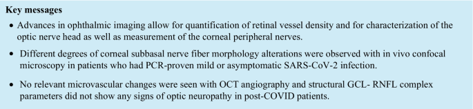
Significantly lower nerve branch density (P = 0.0004), nerve fiber area (P = 0.0001), nerve fiber density (P = 0.0009), nerve fiber length (P < 0.0001), and total nerve branch density (P = 0.002) values were observed in patients after COVID-19 compared to healthy controls. VD of the temporal SCP was significantly different between the two groups (P = 0.019). No other SCP and DCP vessel density parameter differed significantly between the two groups.
Conclusions
Our results suggest that peripheral neurodegenerative changes may occur even after mild or asymptomatic SARS-CoV-2 infection. No relevant microvascular changes were seen with OCT angiography and structural OCT parameters did not show any signs of optic neuropathy in post-COVID patients. In vivo confocal microscopy seems to be an important tool in monitoring peripheral neuropathy in patients after COVID-19.
Open access: https://link.springer.com/article/10.1007/s00417-022-05623-8
Purpose
To examine retinal and corneal neurodegenerative and retinal microvascular changes in patients after mild or asymptomatic COVID-19 disease compared to age-matched controls.
Methods
Thirty-five (35) patients after PCR-proven SARS-CoV-2 infection and 28 age-matched controls were enrolled. Swept-source optical coherence tomography (OCT), OCT angiography, and in vivo corneal confocal microscopy were performed in both groups. Corneal subbasal nerve plexus was quantified. Vessel density for superficial (SCP) and deep capillary plexus (DCP) and structural OCT parameters were recorded.
Results
Significantly lower nerve branch density (P = 0.0004), nerve fiber area (P = 0.0001), nerve fiber density (P = 0.0009), nerve fiber length (P < 0.0001), and total nerve branch density (P = 0.002) values were observed in patients after COVID-19 compared to healthy controls. VD of the temporal SCP was significantly different between the two groups (P = 0.019). No other SCP and DCP vessel density parameter differed significantly between the two groups.
Conclusions
Our results suggest that peripheral neurodegenerative changes may occur even after mild or asymptomatic SARS-CoV-2 infection. No relevant microvascular changes were seen with OCT angiography and structural OCT parameters did not show any signs of optic neuropathy in post-COVID patients. In vivo confocal microscopy seems to be an important tool in monitoring peripheral neuropathy in patients after COVID-19.
Open access: https://link.springer.com/article/10.1007/s00417-022-05623-8