Andy
Senior Member (Voting rights)
Background
In post-COVID-19 conditions (Long COVID), systemic vascular dysfunction is implicated but the mechanisms are uncertain, and treatment is imprecise.
Methods
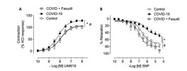
Patients convalescing after hospitalisation for COVID-19 and risk-factor matched controls underwent multisystem phenotyping using blood biomarkers, cardiorenal and pulmonary imaging, and gluteal subcutaneous biopsy (NCT04403607). Small resistance arteries were isolated and examined using wire myography, histopathology, immunohistochemistry, and spatial transcriptomics. Endothelium-independent (sodium nitroprusside) and -dependent (acetylcholine) vasorelaxation and vasoconstriction to the thromboxane A2 receptor agonist, U46619, and endothelin-1 (ET-1) in the presence or absence of a RhoA/Rho-kinase inhibitor (fasudil), were investigated.
Results
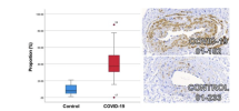
Thirty-seven patients, including 27 (mean age 57 years, 48% women, 41% cardiovascular disease) three months post-COVID-19 and 10 controls (mean age 57 years, 20% women, 30% cardiovascular disease), were included. Compared with control responses, U46619-induced constriction was increased (p = 0.002) and endothelium-independent vasorelaxation was reduced in arteries from COVID-19 patients (p < 0.001). This difference was abolished by fasudil. Histopathology revealed greater collagen abundance in COVID-19 arteries (Masson's Trichrome (MT) 69.7% [95%CI: 67.8, 71.7]; picrosirius red 68.6% [95% CI: 64.4, 72.8]) versus controls (MT 64.9% [95%CI:59.4, 70.3] [p = 0.028]; picrosirius red 60.1% [95% CI: 55.4, 64.8], [p = 0.029]). Greater phosphorylated myosin light chain antibody-positive staining in vascular smooth muscle cells was observed in COVID-19 arteries (40.1%; 95% CI: 30.9, 49.3) vs. controls (10.0%; 95% CI: 4.4, 15.6) (p < 0.001). In proof-of-concept studies, gene pathways associated with extracellular matrix alteration, proteoglycan synthesis, and viral mRNA replication appeared to be upregulated.
Conclusion
Patients with post-COVID-19 conditions have enhanced vascular fibrosis and myosin light change phosphorylation. Rho-kinase activation represents a novel therapeutic target for clinical trials.
Open access, https://academic.oup.com/ehjcvp/advance-article/doi/10.1093/ehjcvp/pvad025/7109259


