Spontaneous, persistent, T cell–dependent IFN-γ release in patients who progress to Long Covid, 2024, Krishna et al

I agree—I think that it actually is unlikely that the state of PEM is a completely unique biological phenomenon in itself. What seems to be ME/CFS-specific is the pattern of being triggered by exertion (as far as we know, we likely can’t confirm that until the mechanism is known).

So there’s:
1) the pathological state that exists when someone is actively in PEM (call it state ‘X’), and
2) the pathway that leads to state ‘X’ in response to overexertion (call it process ‘Y’), and
3) the underlying mechanism which causes a “normal” level of exertion for a healthy person to trigger process ‘Y’ in ME/CFS but not in healthy people

State X is, in all likelihood, not completely unique to ME/CFS. Most people probably experience (at least parts of) it when they have the flu, for example. But it would be useful to have a objective test that can indicate whether or not someone is in state X
 
Last edited:
But it would be useful to have a objective test that can indicate whether or not someone is in state X

People could tell you, though?

There are shades of grey, of course, but if you wanted to run an experiment, it's not difficult for moderately ill to people make sure they're actively in PEM on a particular day.

It's not only quicker than spending years and £££ looking for an objective test, it's potentially just as reliable.
 
People could tell you, though?

I think the idea is to have an objective test for a process. I think we may get into a muddle about terms here but my guess is that jnmaciuch is really suggesting that we need a test for the activity of Y, which will prove objectively that there is a biological cause for being in X when you say you are.

I think tests for both X and Y-->X would be useful in their own regard.

A test for X alone would be useful for the reason that @Sasha already noted--proving that there is actually a biological phenomenon X that is happening when we claim PEM is happening. Ergo, it's not "all in our heads." As you already stated @Jonathan Edwards, if X happens to be similar (or the same, even) as something more societally "legitimate" like viral myalgia, all the better for us.

Primarily, this would be useful on its own for the purposes of any treatment trial. We don't want to rely on subjective reports of PEM because of the heightened susceptibility for confounders.

E.g. if someone just went through one of the "cognitive restructuring" trials which convinced them that PEM isn't real, and then were asked to rate their PEM after the treatment, that would be an invalid measure. But if there is a marker for X, then we could actually check if the participant is still in the state of X after that treatment (even if they claim not to be).

Added: I think this could even be useful as an objective comparative measure for placebo effect for a drug trial. If placebo actually has the power to change something on the relevant biological pathway wrt PEM, a test for X could effectively quantify that.

After that, yes, it would be useful to have a test for the activity of Y leading to X (and that would be where the bulk of useful research happens, in my opinion)
 
Last edited:
What seems to be ME/CFS-specific is the pattern of being triggered by exertion
Add "reliably" to that. There may be people who trigger PEM, but only occasionally, so they don't link exertion with the symptoms. For that matter, maybe some of the symptoms of viral infection are actually from PEM's mechanism, triggered by the immune system's activation.
 
In case it's useful to the IFN-γ discussion:

Single-cell transcriptomics of the immune system in ME/CFS at baseline and following symptom provocation (2024, Cell Reports Medicine)
In particular, gene sets associated with chemokine signaling, migration, and activation are expressed at elevated levels in patient classical monocytes (Figure 2D). Furthermore, we observed the suppression of genes associated with interferon gamma (IFN-γ) signaling in patient classical monocytes. These results suggest that classical monocytes from ME/CFS individuals are biased toward a profile that promotes migration of monocytes to tissue and increased progression toward a macrophage fate. However, we also observed the activation of genes associated with interleukin-10 (IL-10), an anti-inflammatory cytokine. Thus, monocytes in ME/CFS patients may undergo a combination of conflicting inputs.
 
Now that the JE et al hypothesis paper (link to thread) is out, which references this paper, I went back and looked at the figures.

Hypothesis paper said:
T cell responses to peptide antigens are very dependent on contextual signals, and T cells will show apparent ‘autoreactivity’ even in the absence of autoimmune disease.

The relevant T cells may, therefore, also be recognising peptides from similar common ‘junk’ antigens or perhaps with invariant receptors, as for MAIT cells.

They would respond to macrophages that had internalised antigen via FcγRI and presented it together with MHC Class II (or MR1 in the case of MAIT cells) by generating gamma interferon, providing a positive feedback loop with further Fc-gamma-RI expression.

Such a loop might normally serve as a localising, more than an amplification, function – concentrating interacting macrophages and T cells to certain tissue domains – but become persistent in the context of a particular combination of risk factors.

Of interest, a recent study of ‘Long Covid’ cases found increased interferon gamma production from peripheral blood CD8 T cells, dependent on antigen presentation by macrophages

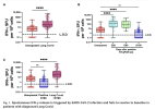
Figure 1 of this paper is quite outstanding, especially in relation to the hypothesis.

Legend of figure
(A) We compared unexposed and Long Covid cohorts
(B) unexposed against each time point for the cohort who experienced acute COVID-19
(C) and then unexposed, acute COVID-19 at day 180 and the Long Covid cohort specifically.
Significance calculated by two-way Kruskal-Wallis analysis of variance (ANOVA), with Dunn’s multiple comparisons test, ****P < 0.0001. ns, not significant.

1752091077223.png
 
Last edited:
I recommend re-visiting this paper again as it is quite fascinating how they determined CD8 cells and macrophages were the ones needed for unstimulated IFN-gamma release in Long Covid.

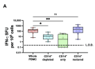
Excerpt for Figure 3
Depletion of CD14+ cells reduced IFN-γ release in all cases. However, the isolated CD14+ cells from the same donor release negligible IFN-γ, suggesting that they are required for IFN-γ production but are not the source of IFN-γ (Fig. 3A).

1752092592838.png
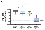
Excerpt for Figure 3C
CD8+ T cells and subpopulations of CD4+ T cells are a major source of IFN-γ. We performed the same cell depletion assays on these populations and found that depletion of CD8+ cells significantly decreased IFN-γ production. A much smaller decrease was noted after CD4+ cell depletion, which was not statistically significant alone but did appear to be additive after CD8+ cell depletion (Fig. 3C).

1752092687648.png
Excerpt from experiment conclusion
Together, our data suggest that CD8+ T cells in contact with CD14+ cells are required for unstimulated IFN-γ production and the majority of this is produced by CD8+ T cells in PBMCs from patients with Long Covid.
 
It looks like this work was done at the University of Cambridge, UK. Does anyone have any contacts with the authors to make them aware of how their work may align with the JE et al hypothesis and try and figure out if CureME biobank ME/CFS, control, and MS samples would be suitable for a replication study. Also, with samples available, they might be interested in extending their work to dig deeper........

What do you think @Jonathan Edwards ?

Also, I wonder if those planning the second round of Stimulate ICP trials are aware of this study...
 
It looks like this work was done at the University of Cambridge, UK. Does anyone have any contacts with the authors to make them aware of how their work may align with the JE et al hypothesis and try and figure out if CureME biobank ME/CFS, control, and MS samples would be suitable for a replication study. Also, with samples available, they might be interested in extending their work to dig deeper........

What do you think @Jonathan Edwards ?

I've just found out that a friend of a friend is part of this group at Cambridge, so I'll ask the friend in the middle to forward the link to JE's Qeios paper.
 
Back
Top Bottom