Proteomic analysis of post-COVID condition: Insights from plasma and pellet blood fractions, 2024, Seco-González et al

forestglip

Senior Member (Voting Rights)
Staff member
Proteomic analysis of post-COVID condition: Insights from plasma and pellet blood fractions

Alejandro Seco-González, Paula Antelo-Riveiro, Susana B. Bravo, P.F. Garrido, M.J. Domínguez-Santalla, E. Rodríguez-Ruiz, Á. Piñeiro, R. Garcia-Fandino

Background
Persistent symptoms extending beyond the acute phase of SARS-CoV-2 infection, known as Post-COVID condition (PCC), continue to impact many individuals years after the COVID-19 pandemic began. This highlights an urgent need for a deeper understanding and effective treatments. While significant progress has been made in understanding the acute phase of COVID-19 through omics-based approaches, the proteomic alterations linked to the long-term effects of the infection remain underexplored. This study aims to investigate these proteomic changes and develop a method for stratifying disease severity.

Methods
Using Sequential Window Acquisition of All Theoretical Fragment Ion Mass Spectra (SWATH-MS) technology, we performed comprehensive proteomic profiling of blood samples from 65 PCC patients. Both plasma and pellet (cellular components) fractions were analyzed to capture a wide array of proteomic changes associated with PCC.

Results

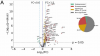
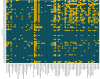
Proteomic profiling revealed distinct differences between symptomatic and asymptomatic PCC patients. In the plasma fraction, symptomatic patients exhibited significant upregulation of proteins involved in coagulation, immune response, oxidative stress, and various metabolic processes, while certain immunoglobulins and proteins involved in cellular stress responses were downregulated. In the pellet fraction, symptomatic patients showed upregulation of proteins related to immune response, coagulation, oxidative stress, and metabolic enzymes, with downregulation observed in components of the complement system, glycolysis enzymes, and cytoskeletal proteins. A key outcome was the development of a novel severity scale based on the concentration of identified proteins, which correlated strongly with the clinical symptoms of PCC. This scale, derived from unsupervised clustering analysis, provides precise quantification of PCC severity, enabling effective patient stratification.

Conclusions

The identified proteomic alterations offer valuable insights into the molecular mechanisms underlying PCC, highlighting potential biomarkers and therapeutic targets. This research supports the development of tailored clinical interventions to alleviate persistent symptoms, ultimately enhancing patient outcomes and quality of life. The quantifiable measure of disease severity aids clinicians in understanding the condition in individual patients, facilitating personalized treatment plans and accurate monitoring of disease progression and response to therapy.

Link | PDF (Journal of Infection and Public Health) [Open Access]
 
The present study extends this inquiry by harnessing the capabilities of SWATH-MS technology, a state-of-the-art data-independent acquisition method that enables a comprehensive and unbiased analysis of the proteome[23]. SWATH-MS is a specific variant of data-independent acquisition (DIA) methods that combines deep proteome coverage capabilities with quantitative consistency and accuracy[24]. This method is not limited by the predefined set of proteins and can detect a wide array of proteins across a vast dynamic range, including low-abundance yet significant biomolecules. SWATH-MS's precision in quantification and identification promises to enhance the detection of subtle proteomic variations and post-translational modifications that remain elusive to conventional binding-affinity based methods.

Notably, we investigate the residual fraction of blood samples (i. e. pellet), a component that has been largely overlooked yet could be instrumental in revealing a subset of proteins not present in plasma, potentially offering new insights into the pathophysiology of PCC (Fig. 1).

I don't know what pellet is. There's not much information online. Maybe this paper has some information:
The second spin step is then performed. ‘g’ for second spin should be just adequate to aid in formation of soft pellets (erythrocyte-platelet) at the bottom of the tube.

From this thread's paper. I think it says RBCs are overrepresented in pellet compared to plasma.

---------

Although both fractions demonstrated correlations between protein profiles and clinical symptoms, the plasma fraction offered a clearer separation, with approximately 85 % agreement between the identified protein clusters and the current clinical symptoms of the patients.

To move beyond the subjective nature of symptom-based classification, the proteins corresponding to the plasma fraction were subjected to an unsupervised k-means cluster analysis, resulting in two well-defined clusters as illustrated in Fig. 2A. Notably, there was an approximate 85 % concordance between symptom-based classifications and the proteomic clusters in the plasma (Table S2), indicating a robust correlation between the plasma plasma proteome and the clinical status of patients.
upload_2024-12-28_16-9-20.png

----
"Volcano plot of differentially expressed proteins in cluster 2 (mostly symptomatic) versus cluster 1 (mostly asymptomatic), using the plasma fraction."
Screenshot from 2024-12-28 16-53-48.png

Upregulated proteins involved in coagulation:
Coagulation Factor XIII B chain (F13B)
Platelet Factor 4 Variant (PF4V)
Fibrinogen chains (FIBA, FIBB, FIBG)
Hemoglobin Subunit Gamma (HBG2)
Antithrombin-III (ANT3)

Upregulated proteins involved in innate immune response:
Properdin (PROP)
C-reactive protein (CRP)
Complement factor H (CFH)
Mannose-Binding Protein C (MBL2)
Haptoglobin (HPT)
Complement C1q subcomponent subunit A (C1QA)
Neutrophil defensin 1 (DEF1)
Protein S100-A9 (S10A9)

Upregulated proteins involved in adaptive immune response:
Immunoglobulin lambda variables 10–54 (LVX54), 6–57 (LV657), 7–43 (LV743), 3–27 (LV327)
Immunoglobulin alpha-2 heavy chain (IGA2)
Immunoglobulin lambda constant 7 (IGLC7)
Immunoglobulin kappa joining 1 (KJ01)
Immunoglobulin heavy variables 3–49 (HV349), 1–18 (HV118), 1–45 (HV145)
Immunoglobulin kappa variables 6–21 (KV621), 1–33 (KV133)

Upregulated proteins involved in oxidative stress response:
Parkinson disease protein 7 (PARK7)
D-dopachrome decarboxylase (DOPD)
Calpain-1 Catalytic Subunit (CAN1)
Calpain Small Subunit 1 (CPNS1)

Upregulated transport proteins
Apolipoprotein C-I (APOC1)
Apolipoprotein B-100 (APOB)
Serotransferrin (TRFE)
Insulin-like Growth Factor-Binding Protein Complex Acid Labile Subunit (ALS)
Hemoglobin Subunit Zeta (HBAZ)

Upregulated proteins related to metabolic processes
ATP-citrate synthase (ACLY)
Transaldolase (TALDO)
Carbonic Anhydrase 1 (CAH1)
Transketolase (TKT)

Upregulated proteins involved in cytoskeletal integrity
Talin-1 (TLN1)
Alpha-actinin-1 (ACTN1)
Drebrin-like protein (DBNL)

Other significant upregulated proteins:
IGH@protein (Q6GMX6)
Aldehyde dehydrogenase family 16 member A1 (A16 A1)
AMBP protein
RD23A
serum amyloid P-component (SAMP)
tropomyosin alpha-4 chain (TPM4)
STIP1
C4BPA
A1AT
KAIN
TCPB
1433E
tetranectin (TETN)
SGTA
FKB1A
URP2
annexin A7 (ANXA7)
ST134
QSOX1
LG3BP
SAA4
APOD
ZA2G
TPM2
LUM
NSF1C
PRDX1
ITIH1
SEPP1
ITIH4
several glycoproteins (A1AG1 and A1AG2).

Downregulated proteins involved in adaptive immune response:
Immunoglobulin heavy constant mu (IGHM)
Immunoglobulin lambda variable 3–10 (LV310)
Immunoglobulin heavy variable 5–10-1 (HV5X1)
Fragment of an immunoglobulin light chain (A0A5C2G2G8)

Other downregulated proteins:
T-complex protein 1 subunit epsilon (TCPE)
14–3-3 protein beta/alpha (1433B)
Glycophorin A (GLPA)
Q8TCD0 (uncharacterized protein)

-----

"Volcano plot of differentially expressed proteins in mild-PCC versus severe-PCC groups using the pellet blood fraction."
upload_2024-12-28_16-56-8.png

distinct patterns of protein expression in both plasma and pellet fractions. Notably, a set of proteins—TRFE, SAMP, TALDO, ACLY, 1433E, MBL2, and ACTN1—were found to be upregulated in symptomatic patients across both fractions. This suggests that these proteins may play crucial roles in the underlying mechanisms of PCC symptoms, given their consistent presence in different blood components. Interestingly, other proteins such as PROP, PARK7, DBNL, FIBA, and FIBB displayed divergent expression patterns: they were upregulated in the plasma fraction but downregulated in the pellet fraction.

Much less skewed using pellet. There is more listing in the paper of proteins involved in various processes as above but for pellet, but I don't want this post to be extremely long.

--------

They made some kind of severity metric using protein levels. As far as I understand, it was made without referencing the group status, just an equation based on the unsupervised clustering results in plasma (A) and pellet (B). This is how the severity metric splits the groups:
upload_2024-12-28_17-13-55.png

--------

Was curious what the difference is between the two types of mannose binding lectin, MBL2 vs MBL1 (Link):
Mannose-binding lectin 1 (MBL1) is a member of the collectin family and is involved in binding multiple types of microorganisms to activate the lectin-complimentary pathway of innate immunity. Most mammals, including rodents and primates have two MBL genes, 1 and 2, however MBL1 has become a pseudogene in the human lineage. Implications for this loss are not understood.
So I guess humans only have MBL2.

--------

Declaration of Generative AI and AI-assisted technologies in the writing process
During the preparation of this work the authors used ChatGPT4o from OpenAI in order to improve language and readability. After using this tool/service, the authors reviewed and edited the content as needed and take full responsibility for the content of the publication.
First time I've seen that in a paper.
 
Last edited:
"Volcano plot of differentially expressed proteins in cluster 2 (mostly symptomatic) versus cluster 1 (mostly asymptomatic), using the plasma fraction."
I think the volcano plots are comparing the two clusters from the unsupervised clustering, not symptomatic vs asymptomatic, which I don't really understand the reason for.

Edit: I guess maybe they're assuming the clustering reveals a subtype of LC?
 
Last edited:
I'm not quite sure how to interpret the volcano plot based on clustering. Whether it's possible the reason so many are upregulated and so few are downregulated is based on clustering, like the algorithm decided something like "all the people with low proteins go in cluster 1, all the high ones go in cluster 2". Maybe someone who understands this can explain.

In any case, I think it said there is 85% overlap between symptom status and clustering.

But anyway, the volcano plot again:
24734-2d6bd66333c7379d2eb1c27c35994c09.jpg

If this study is actually showing that expansive protein upregulation is a feature of long COVID, we've previously seen the same thing. David Price presented that at the PolyBio symposium:
Proteomics: Large numbers of proteins involved in a variety of body systems are upregulated in long COVID.
Screenshot from 2024-12-28 18-54-35.png

I'm not sure if that has been published anywhere. Large numbers of proteins were mostly upregulated in each of four diverse body systems (inflammation, cardiometabolic, neurology, oncology).

He said "This really, I think, reinforces the fact that long COVID is a systemic disease". That doesn't explain why everything is upregulated. If half the significant proteins in neurology were downregulated, neurology could still be involved.

Is there some mechanism that can cause all protein expression to be increased, no matter what it is?

I asked the AI Claude that question. It gave this list. Some or all of this might be wrong, but maybe someone who knows more might see something in here that would fit:
Yes, there are several mechanisms that can lead to broad upregulation of protein expression across multiple systems:
  • Global transcriptional activation:
    • Changes in chromatin structure/accessibility
    • Activation of master transcription factors
    • Epigenetic modifications
  • Enhanced protein synthesis machinery:
    • Increased ribosome biogenesis
    • Upregulation of translation initiation factors
    • mTOR pathway activation (major regulator of protein synthesis)
  • Reduced protein degradation:
    • Decreased proteasome activity
    • Altered autophagy
    • Changes in protein half-life
  • Stress responses:
    • Heat shock response
    • Unfolded protein response (UPR)
    • Cellular stress conditions
  • Hormonal influences:
    • Growth hormone
    • Insulin/IGF-1 signaling
    • Anabolic steroids
  • Inflammation:
    • NF-κB pathway activation
    • Cytokine signaling cascades
 
Last edited:
Hmm, this study comes across as overly definitive and over-confident in the abstract.

A key outcome was the development of a novel severity scale based on the concentration of identified proteins, which correlated strongly with the clinical symptoms of PCC. This scale, derived from unsupervised clustering analysis, provides precise quantification of PCC severity, enabling effective patient stratification.
The quantifiable measure of disease severity aids clinicians in understanding the condition in individual patients, facilitating personalized treatment plans and accurate monitoring of disease progression and response to therapy.

There are no recovered controls, so they avoided major clues that might suggest that some of their findings are just noise.

As for 'asymptomatic PCC', surely that is an oxymoron?
 
Hmm, this study comes across as definitive and over-confident in the abstract.
Probably because ChatGPT partly wrote it, as they disclose.

As for 'asymptomatic PCC', surely that is an oxymoron?
I also was very confused. I just assumed they meant recovered, but I'm not sure. It might mean low number of symptoms.

Figure 3C has the reported symptoms of each patient:
upload_2024-12-28_19-43-17.png

Hard to read the symptoms, but there are some participants with no symptoms. (Edit: Though by my count there are only 5 participants with no symptoms out of 65.)
 
Last edited:
Proteomic and metabolomic profiling of plasma uncovers immune responses in patients with Long COVID-19, 2024, Wei et al

This preprint did untargeted proteomics as well.
Compared to the HC group, the LC group had 187 proteins upregulated and 144 proteins downregulated.

In comparison to the HC group, the Recovered group had 93 proteins upregulated and 65 proteins downregulated.

When comparing the LC group to the Recovered group, 113 proteins were upregulated, and 86 proteins were downregulated.

Screenshot from 2024-12-28 20-01-25.png

Not as striking as the previous two studies I referenced, but still more proteins are upregulated than downregulated when comparing LC to never infected (56%) or LC to recovered (57%).
 
Last edited:
As for 'asymptomatic PCC', surely that is an oxymoron?

I haven't read this yet, but it may mean "not yet symptomatic" - ie like a cardiovascular or diabetic risk profile before angina/MI or DM declares months/years later.

Their previous paper is Lipidomics signature in post-COVID patient sera and its influence on the prolonged inflammatory response (2024, Journal of Infection and Public Health)

Edit: no this was a prospectively tracked cohort of 65 patients. They had blood samples taken at a single time-point (variable between the patients) following confirmed infection (non-hospitalised). The term "PCC" seems to be being used as the condition of being post-Covid, so some are symptomatic and some asymptomatic at the time of their blood sample. They do look at patient status: at 90 days, 9 months; and at sample collection. Some patients had no symptoms at 9 months.
 
Last edited:
This study did untargeted proteomics, but on extracellular vesicles not plasma:

Dysregulation of extracellular vesicle protein cargo in female ME/CFS cases & sedentary controls in response to maximal exercise, 2023 Giloteaux et al

upload_2024-12-28_20-42-35.png

Split evenly at baseline, most proteins downregulated at 15 minutes post-exercise, maybe slightly more upregulated at 24 hours post-exercise.

I found this interesting though:
upload_2024-12-28_20-44-5.png

These are separate volcano plots for ME/CFS and controls. Each one shows the level of proteins in the group 15 minutes after exercise compared to before exercise. In both groups there is widespread upregulation of proteins in EVs due to exercise.
 
Data-independent LC-MS/MS analysis of ME/CFS plasma reveals a dysregulated coagulation system, endothelial dysfunction, downregulation of complement machinery, 2024, Nunes, Kell, Pretorius et al

This one did proteomics on plasma, like the first three studies, but on ME/CFS. Slightly more proteins upregulated than downregulated in ME/CFS (53% vs 47%), but nothing special.
Our experiment indicates that 24 proteins are significantly increased in the ME/CFS group compared to the controls, and that 21 proteins are significantly downregulated.
 
I also was very confused. I just assumed they meant recovered, but I'm not sure. It might mean low number of symptoms.

Figure 3C has the reported symptoms of each patient:


Hard to read the symptoms, but there are some participants with no symptoms. (Edit: Though by my count there are only 5 participants with no symptoms out of 65.)

Oh, one of the "symptoms" listed is "Symptomatic". There are 8 not filled in for this. None of them have any long COVID symptoms. 3 have boxes filled in for medications or unrelated symptoms like high cholesterol.
 
Proteins with more than 10 peptides and seven transitions were selected for quantification. Any shared or modified peptides were excluded.
If anyone is familiar with the technique used to identify the proteins and feels inclined to describe it here, that would be great.


Proteins exhibiting zero variance across all patients were eliminated, resulting in the exclusion of 21 proteins from the plasma sera samples, leaving 294 remaining. Subsequently, a power transformer (using the Yeo- Johnson method)[36] was applied to adjust the protein concentra- tions towards a more normal distribution. Following the same pre- processing protocol, the pellet fraction retained all 315 proteins, as no proteins were eliminated in this fraction.
They then did a PCA analysis and found that the PCA analysis of the sera divided the participants into two groups, one with mostly participants asymptomatic at the time of blood draw, and one with participants mostly symptomatic. However, I note that PC1 only accounted for 11% of the variation. The PCA analysis for the pellet is reported to be less useful at dividing participants. It is the two groups identified by PC1 for the sera samples that are used in subsequent volcano plots.

Notably, these clusters showed an 85 % correspondence with clinical symptomatology at the time of the blood extraction, where one cluster predominantly consisted of symptomatic patients and the other of asymptomatic patients, as detailed in Table S2.
I don't know what clinical symptomatology at the time of the blood extraction was considered. I think it is the Patient Status at Sample Extraction i.e.
Physical deconditioning
Cough and/or throat clearing
Anosmia and/or ageusia
Difficulty concentrating
Insomnia
Headache
Myalgias
Alopecia​


Moreover, to quantify the severity of illness for each patient, we proposed a severity metric model (S) that depends solely on the protein concentrations and the identified clusters.
Just noting here that the 'severity metric' is not based on the actual severity of the illness the participants have. It is based on how well the participant's protein levels match those of the two groups ("mostly asymptomatic", "mostly symptomatic") identified in the PCA. I think that 'severity metric' is a misleading name.

PC1 does give a pretty impressive separation of the people who ticked one or more of the 'symptom' boxes at blood collection time. But, with 'physical deconditioning' being a symptom, it does raise the question as to how much of the different protein levels are due to differences in activity levels.
 
Last edited:
Yeah, unfortunately it's a bit questionable. For example:

Participant 2 had physical deconditioning, anosmia/ageusia and difficulty concentrating when they gave the sample. But they are categorised as being in the 'asymptomatic' group.

Participant 4 only ticked the symptom box for physical deconditioning when they gave the sample, but has high cholesterol, is obese and has ischaemic heart disease, and is categorised in the 'symptomatic' group.

Participant 23 has connective tissue disease, and is categorised in the 'symptomatic' group.

I don't think the characterisation of the post-Covid condition is good enough to warrant the analysis of the proteins. It's really tempting to sift through the proteins they identified as different, but I don't think they will tell us anything reliable about post-Covid-19 ME/CFS.

I like the technology and I hope the team do more proteomic analyses of PCC, but they need to characterise their participants much more stringently.
 
Last edited:
"Volcano plot of differentially expressed proteins in cluster 2 (mostly symptomatic) versus cluster 1 (mostly asymptomatic), using the plasma fraction."
View attachment 24783

That's unbalanced enough that despite the label I'm wondering if they've actually remembered to log transform on the x-axis. (A protein can rise by more than 100% but not fall by more than 100%)?!
 
Back
Top Bottom