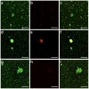
Plasma from patients with pulmonary embolism show aggregates that reduce after anticoagulation
Baker, Stephen R.; Halliday, Georgia; Ząbczyk, Michal; Alkarithi, Ghadir; Macrae, Fraser L.; Undas, Anetta; Hunt, Beverley J.; Ariëns, Robert A. S.
Background
Microclots, a term also used for amyloid fibrin(ogen) particles and henceforth named aggregates, have recently been reported in the plasma of patients with COVID-19 and long COVID. These aggregates have been implicated in the thrombotic complications of these diseases.
Methods
Plasma samples from 35 patients with acute pulmonary embolism were collected and analysed by laser scanning confocal microscopy and scanning electron microscopy before and after clotting.
Results
Here we confirm the presence of aggregates and show that they also occur in the plasma of patients with pulmonary embolism, both before and after clotting. Aggregates vary in size and consist of fibrin and platelets. We show that treatment with low-molecular weight heparin reduces aggregates in the samples of patients with pulmonary embolism. Double centrifugation of plasma does not eliminate the aggregates.
Conclusions
These data corroborate the existence of microclots or aggregates in diseases associated with venous thromboembolism. Important questions are raised regarding their pathophysiological relevance and further studies are warranted to investigate whether they represent cause or consequence of clinical thrombosis.
Plain Language Summary
When blood turns from liquid to solid, a protein called fibrin and cells called platelets aggregate to form a blood clot. Small aggregates have been found in the blood of people with COVID-19 and long COVID. Here, we show that small aggregates also occur in the blood of patients with pulmonary embolism, a disorder in which blood clots are trapped in an artery in the lung, preventing blood flow. We confirm that aggregates consist of fibrin and platelets, and show that the number of aggregates is lower when patients are treated with blood thinning drugs. These results suggest other disorders of the blood should also be investigated to see whether aggregates are present and whether they have an impact on the outcome for the patient. This could help us understand the cause of diseases associated with blood clotting, which might offer new approaches for diagnosis and treatment.
Link | PDF (Nature Communications Medicine)

