Immunological and Clinical Markers of Post-acute Sequelae of COVID-19: Insights from Mild and Severe Cases 6 Months Post-infection, 2025, Mouton et al

SNT Gatchaman

Senior Member (Voting Rights)
Staff member
Immunological and Clinical Markers of Post-acute Sequelae of COVID-19: Insights from Mild and Severe Cases 6 Months Post-infection
William Mouton; Sophia Djebali; Marine Villard; Omran Allatif; Cécile Chauvel; Sarah Benezech; Philippe Vanhems; Jacqueline Marvel; Thierry Walzer; Sophie Trouillet-Assant

Post-acute sequelae of COVID-19 (PASC) are a complex clinical condition that requires a better understanding of its underlying biological mechanisms.

In this study, we assessed hundreds of virological, serological, immunological, and tissue damage biomarkers in two cohorts of patients who had experienced either mild (n = 270) or severe (n = 188) COVID-19, 6 to 9 months post-initial infection, and in which 40% and 57.4% of patients, respectively, developed PASC.

Blood analysis showed that the main differences observed in humoral, viral, and biological biomarkers were associated with the initial COVID-19 severity, rather than being specifically linked to PASC. However, patients with PASC displayed altered CD4+ and CD8+ memory T cell subsets, with higher cytokine-secreting cells and increased terminally differentiated CD45RA+ effector memory T cells (TEMRA). Elevated SARS-CoV-2-specific T cells responsive to nucleocapsid/membrane proteins with a TEMRA phenotype were also observed. A random forest model identified these features and initial symptom duration as top variables discriminating PASC, achieving over 80% classification accuracy.

Link | PDF | European Journal of Immunology [Open Access]
 
No evidence of anti-interferon auto-antibodies —

We […] observed a low prevalence of anti-IFN-α2 plasmatic autoantibodies with neutralizing capacity; among the 458 patients, they were found in only six severe patients (1.3%) without association with PASC (data not shown).

No evidence for viral persistence —

Regarding circulating SARS-CoV-2 nucleocapsid antigens, they were not detectable in any of the samples.

No evidence for latent viral reactivation —

Similarly, cytomegalovirus (CMV) and adenovirus were not detectable, while only 10 samples were positive for Epstein–Barr virus (EBV; 2.2%), without association with PASC.

Torque teno virus positivity correlated with PASC, though viral load did not correlate —

Conversely, torque teno virus (TTV) load was detectable in most samples and was significantly different between cohorts; severe patients had a higher viral load than mild patients (3.05 [2.23–3.73] vs. 2.43 [1.80–3.00] Log 10 copies/mL, p < 0.0001). However, while a higher proportion of TTV-positive samples was found in severe patients with PASC compared with those without (58% vs. 42%, p = 0.0068), no significant difference in TTV loads was observed according to the PASC status

No difference in cortisol —

cortisol was quantified in 160 of the available 175 samples (with enough volume), and significantly higher levels were observed in severe patients, as compared with mild patients (90.9 [73.9–124.4] vs. 74.9 [60.9–101.4] pg/mL, p = 0.0045). However, no significant difference in cortisol levels was found between patients with and without PASC, neither in the mild (93.6 [63.4–109.2] vs. 71.2 [55.3–93.4] pg/mL, p = 0.070) nor in the severe cohort (90.4 [67.6–125.3] vs. 100.3 [80.4–118.3] pg/mL, p = 0.38).
 
Last edited:
On immune cell subsets —

patients with PASC tended to exhibit higher counts of lymphocytes, while patients without PASC tended to exhibit higher counts of total and classical monocytes, IgD + CD27 + B cells, and CD45RA − /CD95 + CD8 + T cells. These results highlight notable alterations in T cell counts, which were previously reported in patients with PASC36]; therefore, we performed unsupervised clustering of T cell data

On T cell markers —

Several markers were differentially expressed between the two groups, mainly in memory CD4+ and CD8+ T cells. Among memory CD4+ T cells, effector memory T cells re-expressing CD45RA (TEMRA, cluster 17) and effector memory T cells (TEM, cluster 10) were observed to have a similar abundance but distinct phenotypes depending on the PASC status.

In patients with PASC, CD4+ TEMRA cells (cluster 17) showed higher expression of CD57 (p = 0.015) and Tbet (p = 0.048) as well as lower CXCR3 expression (p = 0.015). Furthermore, CD4 + TEM cells (cluster 10) in these patients showed reduced CD27 expression (p = 0.026).

Regarding memory CD8+ T cells, despite a similar abundance found among patients with and without PASC (Figure 4D), CD8 + TEMRA cells (cluster 5) from patients with PASC displayed higher Tbet expression (p = 0.032) and lower CXCR3 expression (p = 0.0072).

On response to viral antigens —

The response to S was similar in both groups, but patients with PASC exhibited a significantly higher response to N/M-derived peptides
 
ML analysis (Random Forest) —

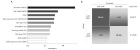
In order to find the variables that best discriminated patients with PASC from those without, we used a random forest algorithm as well as the 674 clinical, serological, virological, and immunological parameters monitored in this study. […] top 10 and top 50 discriminant parameters. This list included clinical (symptom duration, weight), plasmatic (GFAP, IL-6, Nf-L, and IFN-λ 1 ), as well as immunological parameters (CD8 + TEMRA CXCR3, CD4 + TEMRA Ki67, N/M-responsive CD4 + TEMRA, classical monocytes).

No single parameter distinguished —

The most important parameter identified was the symptom duration, followed by several immune parameters associated with T cells. However, none of these parameters alone was sufficient to fully differentiate patients with PASC from those without.

Top 10 parameters —

We then assessed the capacity of the top 10 parameters to correctly discriminate altogether patients with and without PACS, using a confusion matrix. As shown in Figure 6B, these 10 parameters had a classification performance greater than 80% for both groups of patients, supporting a role for T cells in PASC.

Classifier.jpg

  • Symptom duration
  • CD8 TEMRA CXCR3
  • GFAP
  • N/M-responsive CD4+ CXCR3 CD57
  • CD4 Naive PD1
  • CD4 TEMRA Ki67
  • CD4 Tregs CD39+ Ki67
  • CD4 Naive CD57
  • Body weight
  • N/M-responsive CD4+ TEMRA
 
Last edited:
Back
Top Bottom