Hamsters with long COVID present distinct transcriptomic profiles associated with neurodegenerative processes in brainstem 2025 Coleon et al.

Jaybee00

Senior Member (Voting Rights)
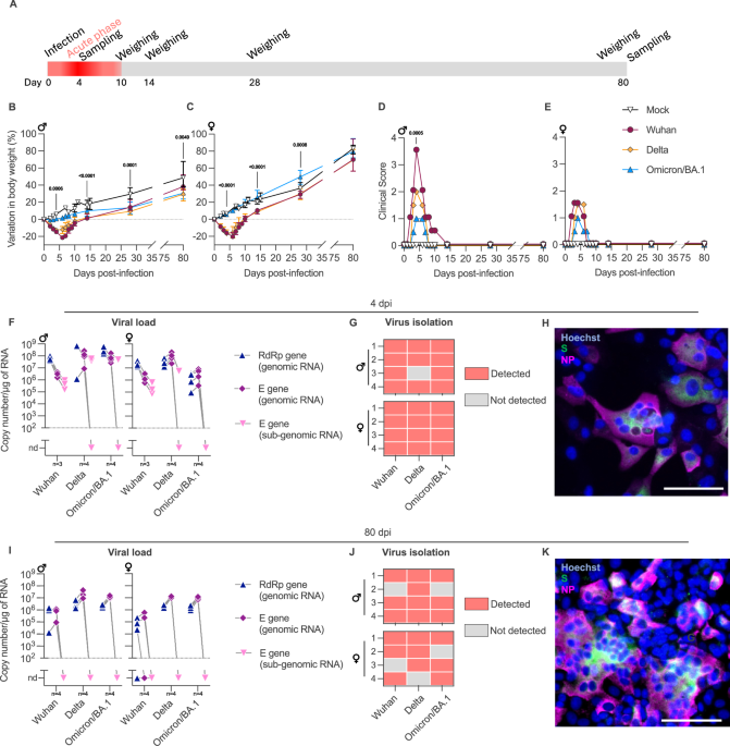
Following infection with SARS-CoV-2, patients may experience with one or more symptoms that appear or persist over time. Neurological symptoms associated with long COVID include anxiety, depression, and memory impairment. However, the exact underlying mechanisms are not yet fully understood. Using golden hamsters as a model, we provide further evidence that SARS-CoV-2 is neuroinvasive and can persistently infect the brain, as viral RNA and replicative virus are detected in the brainstem 80 days after the initial infection. Infected hamsters exhibit a neurodegenerative signature in the brainstem, characterized by overexpression of innate immunity genes, and altered expression of genes involved in the dopaminergic and glutamatergic synapses, in energy metabolism, and in proteostasis. These infected animals exhibit persistent depression-like behavior, impaired short-term memory, and late-onset signs of anxiety. Finally, we provide evidence that viral and immunometabolic mechanisms coexist in the brainstem of SARS-CoV-2-infected hamsters, contributing to the manifestation of neuropsychiatric and cognitive symptoms.


I feel sorry for the hamsters.

As an immune induced neurological syndrome though this is a much better model of longcovid and even ME than mouse models based on exhaustion from swimming.

To make it more relevant they need to take observations from the hamster model and then make predictions and look for evidence of similar in humans with longcovid. If some do and some don't (resemble hamster physiology) then we need to start subtyping longcovid along those lines.
 
We then investigated the presence and persistence of the different SARS-CoV-2 variants in the brainstem of intranasally-inoculated hamsters. To this aim, we initially performed a follow-up study to quantify the viral load in the brainstem at different time-points after infection of male and female hamsters with SARS-CoV-2 Wuhan. Remarkably, genomic and sub-genomic viral RNA were detected in the brainstem as early as four hours after the intranasal inoculation, with stable and elevated levels at 1-, 2- and 4-dpi, regardless of the sex. Genomic RNA was still detected at 14-, 30- and 80-dpi, but in contrast, sub-genomic RNA was below the limit of detection (Supplementary Fig. 2A, C)

So, there's this interesting finding of genomic RNA persisting in the brain stem, but sub-genomic RNA not. These charts are from the Supplementary Materials. Chart A is for males, Chart B is for females. The pink triangles are the sub-genomic RNA. It looks like for some hamsters, sub-genomic RNA was not found throughout the time series. But, after 14 days, no hamsters had sub-genomic RNA in the brain stem, whereas genomic RNA was.

I'm not sure what to make of that. Subgenomic RNA seems to be just fragments of RNA, although intentionally made, which I think can help a virus evade pathogen recognition mechanisms of the host. I'll be interested to see what the authors make of that finding in the discussion.
 
Last edited:
Remarkably, we still detected viral genomic RNA in the brainstem of the animals at 80 dpi (Fig. 1I). Although sub-genomic RNA was below the limit of detection, indicating low viral load, we were able to isolate and amplify infectious SARS-CoV-2 from the brainstem of infected hamsters at 80 dpi (Fig. 1K, Supplementary Fig. 3B). In these samples, the viral titers were remarkably low (<102 TCID50/mL), yet we observed cytopathic effect and detected viral proteins five to seven days after infection of the cells.
We amplified infectious virus from the brainstems of 75% of Wuhan-infected animals (3/4 males and 3/4 females), 87.5% of Delta (4/4 males and 3/4 females), and 75% of Omicron/BA.1 (3/4 males and 3/4 females) at 80 dpi (Fig. 1J). In parallel, we also quantified the viral RNA loads in the airways (nasal turbinates and lungs) and in other parts of the brain than the brainstem (olfactory bulbs, cerebral cortex, cerebellum) of the animals at 80 dpi. Genomic SARS-CoV-2 RNA was detected in the airways of all animals. In the other brain areas, however, the detection was variable (Supplementary Fig. 4), and no sub-genomic viral RNA was detected.
So, they found low levels of viral RNA in a lot of places, the brain stem, nasal turbinates, lungs, and even other parts of the brain at 80 days.

Having demonstrated the persistence of SARS-CoV-2 RNA and infectious virus in the brainstem of infected hamsters, we next analyzed the histopathological changes at 4 and 80 dpi, focusing on the olfactory bulbs (considered the entry point of the virus into the brain) and on the brainstem (Fig. 2A–C). In general, the histopathological analysis of the brain revealed no relevant microscopic alterations neither during the acute nor during the late phase of the infection (Supplementary Fig. 5).
So, despite the presence of the RNA, they didn't find structural changes.
We also quantified the levels of serum neurofilament light chain (NfL) to assess neuroaxonal damage in male and female hamsters intranasally-inoculated with Wuhan at 80 dpi, but observed no difference between infected and mock-infected animals (Supplementary Fig. 2B, D).
And they didn't find NfL, a marker of neuron damage.
 
But, they did find higher levels of GFAP (glial fibrillary acidic protein) - they say this is indicative of astrogliosis.

Figure 2

Screenshot 2025-07-23 at 3.28.43 pm.png


They also looked at the brainstem transcriptome at 80 days (infected group versus sham infected group). There are pathways that we have seen come up as important in human Long Covid - neurons, synapses, energy.
During this late phase of SARS-CoV-2 infection, the dysregulated biological processes in the brainstem (234 GO terms) mainly involved energy, synapses, axonogenesis and neurogenesis; the cellular component (131 terms) was identified mainly as targeting synaptic zones and mitochondria; and molecular function (28 GO terms) was related to energy and calcium activity (Fig. 3)
Further, twenty-three KEGG pathways were significantly regulated, mostly related to neurodegeneration, neurotransmission, calcium signaling and energy (Fig. 4A).
 
But, did these hamsters really have Long Covid? and a Long Covid that is like ME/CFS?

we decided to test whether infected animals also exhibit clinical manifestations related to long Covid, specifically anxiety, depression, and memory loss, which may be related to neurodegenerative processes. To this end, we performed three well-described and recognized behavioral tests to assess anxiety, depression, and recognition memory in the animals. The hamsters performed four sequential behavioral tests (one test/day) on three sessions: the first session just after the acute phase (between 14-17 dpi, hereinafter called 15 dpi session), the second between 28-31 dpi (hereinafter called 30 dpi session), and the third session between 76-79 dpi (hereinafter called 80 dpi session) (Fig. 6A). To properly quantify the effect of covariates (sex, time post-infection, SARS-CoV-2 variant), we used mixed model regression to account for within-individual correlation arising from repeated measurements.

Screenshot 2025-07-23 at 4.15.07 pm.png

I'm rather doubtful that these tests mean what the authors think they mean. The results aren't quite a coherent story, but, still, there might be something there. The results of some tests are relegated to the Supplementary Materials.

For example, the middle charts are a sugar splash, with the amount of time then spent grooming recorded.
Time grooming corresponds to an index of motivational and self-care behavior and decreased grooming time is considered as a depression-like behavior.
Reduced grooming time might also be related to fatigue. The grey band is the range of times in mice that had the mock infection. The top charts are for male hamsters, the bottom charts are for female hamsters. The different lines are for different strains of the SARS-CoV-2 virus. Female hamster that were infected did seem to spend less time grooming than the uninfected hamsters.

We provide conclusive evidence that neuropsychiatric symptoms and cognitive impairment related to long COVID follow the acute infection, in a model without social or somatization influence, and no effects related to post-intensive care syndrome.
I think that claim is a bit of a stretch. But, it's a paper that I think is worth a read.

These cognitive and neuropsychiatric symptoms can have different origins and causes; though, the asset of the hamster model is that the behavioral tests took place in an environment free from social pressure or risk of somatization, i.e., infected and mock-infected animals (both male and female) were tested simultaneously under the exact same conditions. Consequently, the differences observed in clinical profile between the infected and mock-infected animals can only be explained by the SARS-CoV-2 infection itself, and the underlying mechanisms are likely to be associated with virus-related and neuro-immunometabolic changes in their brainstem.
 
Last edited:
Golden hamsters are a commonly used animal model to study COVID. You will find them used as a robust preclinical model in a lot of studies https://www.pnas.org/doi/10.1073/pnas.2009799117
Sure, they’re commonly used. But there is little evidence we understand what long covid is or that animal models are particular useful for transferring these ideas between humans and animals. I’m not discounting their use in understanding disease but I am extremely sceptical of the way in which direct comparisons like this are made and stated as fact in many papers which use them.
 
Back
Top Bottom