Kalliope
Senior Member (Voting Rights)
Transl Med
Endothelial dysfunction and altered endothelial biomarkers in patients with post-COVID-19 syndrome and chronic fatigue syndrome (ME/CFS) Milan Haffke et al
Abstract
Background: Fatigue, exertion intolerance and post-exertional malaise are among the most frequent symptoms of Post-COVID Syndrome (PCS), with a subset of patients fulfilling criteria for Myalgic Encephalomyelitis/Chronic Fatigue Syndrome (ME/CFS). As SARS-CoV-2 infects endothelial cells, causing endotheliitis and damaging the endothelium, we investigated endothelial dysfunction (ED) and endothelial biomarkers in patients with PCS.
Methods: We studied the endothelial function in 30 PCS patients with persistent fatigue and exertion intolerance as well as in 15 age- and sex matched seronegative healthy controls (HCs). 14 patients fulfilled the diagnostic criteria for ME/CFS. The other patients were considered to have PCS. Peripheral endothelial function was assessed by the reactive hyperaemia index (RHI) using peripheral arterial tonometry (PAT) in patients and HCs. In a larger cohort of patients and HCs, including post-COVID reconvalescents (PCHCs), Endothelin-1 (ET-1), Angiopoietin-2 (Ang-2), Endocan (ESM-1), IL-8, Angiotensin-Converting Enzyme (ACE) and ACE2 were analysed as endothelial biomarkers.
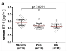
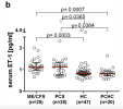
Results: Five of the 14 post-COVID ME/CFS patients and five of the 16 PCS patients showed ED defined by a diminished RHI (< 1.67), but none of HCs exhibited this finding. A paradoxical positive correlation of RHI with age, blood pressure and BMI was found in PCS but not ME/CFS patients. The ET-1 concentration was significantly elevated in both ME/CFS and PCS patients compared to HCs and PCHCs. The serum Ang-2 concentration was lower in both PCS patients and PCHCs compared to HCs.
Conclusion: A subset of PCS patients display evidence for ED shown by a diminished RHI and altered endothelial biomarkers. Different associations of the RHI with clinical parameters as well as varying biomarker profiles may suggest distinct pathomechanisms among patient subgroups.


