Effects of Pseudoserum on Thrombin-Induced Fibrin Networks: Potential Clinical Insight into Coagulation Independent of Clotting Factors, 2025, Nunes

Mij

Senior Member (Voting Rights)

Abstract​

Coagulation, although primarily regulated by platelets, endothelial cells, and clotting factors, can also be influenced by molecules that are not traditionally seen as related to coagulation, including cytokines, hormones, metabolites, reactive oxygen species, acute phase reactants, and more.

Here, we derive pseudoserum or clotting factor-depleted fractions from control, type II diabetes mellitus, and Long COVID platelet-poor plasma (PPP) samples, and expose them to purified, exogenous fibrinogen obtained from healthy donors. Thrombin-induced fibrin networks were then formed and visualized using light and scanning electron microscopy.

The results demonstrate that pseudoserum can greatly influence the organisation, density, and ultrastructure of fibrin networks formed from purified fibrinogen, emphasizing the role of non-clotting factors in fibrin formation. Fibrin networks formed from purified fibrinogen exposed to control pseudoserum appear homogeneous, exhibiting organized architecture with few regions of unusual density or aggregates, whereas the networks formed using patient pseudoserum show disorganisation, regions of density, fibre-like strands, and anomalous aggregates.

These abnormalities are also observed in patient PPP samples, suggesting that fibrin network characteristics in PPP samples are also significantly influenced by non-clotting factors and are somewhat independent of endogenous fibrinogen. The ability of pseudoserum to drive these changes, despite the absence of endogenous fibrinogen and other classical clotting factors, suggests that soluble molecules retained in pseudoserum can directly modify fibrinogen’s structural conformation and functionality, influence thrombin-mediated fibrin formation and polymerization, and/or impact Factor XIII’s crosslinking capabilities.

This study provides a systems-level perspective on the influence of pseudoserum on fibrin networks and highlights the potential of serum and other clotting factor–depleted fractions to yield deeper mechanistic and diagnostic insights into coagulation.
LINK
 
My notes of the main ideas presented —

Fibrinogen is made by the liver and is soluble. With thrombin it forms initially small fibrin monomers (variably soluble to insoluble) which then aggregate to form a larger scaffold, which with platelets forms the typical clot.

Apart from the timing to formation, it is established that the structure of the fibrin scaffold is dependent on clotting factors ("thickness, branching, pore size, and susceptibility to lysis").

Also non-clotting factors modulate this process —

A number of these molecules, including proinflammatory cytokines, serum amyloid A, reactive species, hormones, and sugars, can directly interact with and alter the structure and function of fibrinogen, which can influence resulting clot networks, including fibre thickness, pore size, and resistance to fibrinolysis

You can look at serum (what remains after removing the clotted component of blood) or plasma (blood with its clotting factors still present, but inactivated). You can also remove platelets from plasma: platelet-poor plasma (PPP); platelet-rich plasma (PRP).

"Due to the fact that serum mostly lacks fibrinogen and other clotting factors, and hence cannot coagulate, serum is typically viewed to lack clinical utility in the context of coagulation."

What they're looking at is the effect of healthy vs disease serum when you add back in healthy fibrinogen and see what the fibrin structure is. They refer to this as "pseudoserum".

They made the serum by clotting existing PPP samples (from their prior studies) adding calcium in the thromboelastography machine, and keeping the unclotted residual. They then formed pseudoserum by adding healthy purified fibrinogen. Then they added purified thrombin to form fibrin.

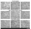
Yann04's images above show the difference in the fibrin scaffold structures (fig 2).

There are clear morphological distinctions between control and disease fibrin networks, with the latter showing enhanced density and disorganisation, dense granular regions, fibre thickness, and anomalous deposits. The control fibrin clots, in contrast, exhibit structural uniformity, almost lack regions of granular densities, and exhibit notable pore sizes and sparsely arranged networks.

They conclude that this pseudoserum model can be used to evaluate the known non-clotting factor modulators of clotting —

The ability of pseudoserum to drive these changes, despite the absence of fibrinogen and other clotting factors, emphasizes that soluble molecules retained in pseudoserum can modulate the coagulation process.

Together, these data reposition pseudoserum and other clotting-factor–depleted fractions as a practical model to study serum-driven influences on fibrin architecture, expanding the biology of fibrin modulation and flagging serum-based assays as promising adjunct diagnostic and prognostic tools for coagulopathies.
 
The paper's comments on amyloid microclots

Fibrin networks were also stained with ThT to assess amyloid areas in fibres and also to determine if fibrinaloid microclot complexes are structurally associated or embedded within the fibrin networks. However, no signal was observed within fibrin fibres or networks. Hence it seems that pseudoserum exposure to purified fibrinogen did not result in noticeable amyloid or fibrinaloid microclot complexes that are embedded in and structurally associated with the induced fibrin network.

While no signal was observed within the fibrin network, there were some ThT-positive aggregates that appeared on top, under, and next to the networks, which we propose represent those already present in pseudoserum (Figure 3).

Additionally, exposing pseudoserum to purified fibrinogen did not result in the formation of ThT-positive aggregates incorporated within fibrin networks. Instead, the aggregates already present in pseudoserum appeared excluded from further fibrin polymerization, even in the presence of exogenous fibrinogen and thrombin[…] It is also notable that many aggregates in pseudoserum did not stain with ThT, suggesting the presence of a distinct non-amyloid fraction that still appears more prevalent in disease groups.

we found that fibres formed by adding patient pseudoserum to purified fibrinogen retained a dense matted appearance, but these structures did not stain with ThT and were therefore not amyloid. This distinction suggests that some amyloid deposits represent insoluble fibrinogen that has misfolded into amyloid and become associated with pathological cellular debris and neutrophil extracellular traps (NETs), forming what we now define as fibrinaloid microclot complexes. In contrast, residual soluble fibrinogen in circulation may still polymerize into dense matted fibrin fibres that are structurally abnormal but not amyloid.

Together, these findings reveal two distinct but co-existing pathological processes: (1) pre-formed amyloidogenic deposits in circulation, and (2) soluble fibrinogen that polymerizes into non-amyloid dense matted fibrin fibres.
 
Back
Top Bottom