You are using an out of date browser. It may not display this or other websites correctly.
You should upgrade or use an alternative browser.
You should upgrade or use an alternative browser.
Community Symposium on the Molecular Basis of ME/CFS Sept 5 (Stanford/Ron Davis)
- Thread starter Jaybee00
- Start date
No I must have misremembered if they're saying they plan to add more males.But I've been knocked flat by a new med, so my brain's all over the place—apologies if I misunderstood.
The tracers are certain molecules that release radiation. If they're inside a patient, a PET scanner can detect the radiation which lets you pinpoint the exact location it came from, thus where in the body the tracer is.By the way, I am a bit fuzzy on exactly what these tracers do and precisely what it is they measure.
Would anyone care to explain like I'm brain foggy? (because I am!)
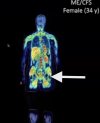
They make tracers that stick to specific molecules that exist inside humans that they are interested in finding. In the above case, the tracer 11C-DPA-713 sticks to a protein called TSPO that some cells have. If you inject the tracer, it sticks to the TSPO in the body, wherever it happens to be, and then lights up on the scanner images showing you where the TSPO is.
So the images are basically, where is the TSPO in each person, and how much is there.
And hopefully someone else can provide more detail about TSPO specifically, but just going by what she said in the symposium and research roadmap webinar, it's a molecule on a cell's mitochondria. Some of the cells that have the highest levels of it are glial cells and immune cells, maybe highest of all being microglia and macrophages but I'm not positive about that. So if you see a lot of TSPO in a certain location, that might mean there are lots of immune cells there for some reason.
V.R.T.
Senior Member (Voting Rights)
Thanks, that's really helpful!The tracers are certain molecules that release radiation. If they're inside a patient, a PET scanner can detect the radiation which lets you pinpoint the exact location it came from, thus where in the body the tracer is.
They make tracers that stick to specific molecules that exist inside humans that they are interested in finding. In the above case, the tracer 11C-DPA-713 sticks to a protein called TSPO that some cells have. If you inject the tracer, it sticks to the TSPO in the body, wherever it happens to be, and then lights up on the scanner images showing you where the TSPO is.
So the images are basically, where is the TSPO in each person, and how much is there.
And hopefully someone else can provide more detail about TSPO specifically, but just going by what she said in the symposium and research roadmap webinar, it's a molecule on a cell's mitochondria. Some of the cells that have the highest levels of it are glial cells and immune cells, maybe highest of all being microglia and macrophages but I'm not positive about that. So if you see a lot of TSPO in a certain location, that might mean there are lots of immune cells there for some reason.
V.R.T.
Senior Member (Voting Rights)
So could one conceivably make tracers for gamma delta t cells, gamma/type 1 interferons, or whatever other cells we might suspect are hiding from other methods of detection?The tracers are certain molecules that release radiation. If they're inside a patient, a PET scanner can detect the radiation which lets you pinpoint the exact location it came from, thus where in the body the tracer is.
They make tracers that stick to specific molecules that exist inside humans that they are interested in finding. In the above case, the tracer 11C-DPA-713 sticks to a protein called TSPO that some cells have. If you inject the tracer, it sticks to the TSPO in the body, wherever it happens to be, and then lights up on the scanner images showing you where the TSPO is.
So the images are basically, where is the TSPO in each person, and how much is there.
And hopefully someone else can provide more detail about TSPO specifically, but just going by what she said in the symposium and research roadmap webinar, it's a molecule on a cell's mitochondria. Some of the cells that have the highest levels of it are glial cells and immune cells, maybe highest of all being microglia and macrophages but I'm not positive about that. So if you see a lot of TSPO in a certain location, that might mean there are lots of immune cells there for some reason.
Last edited:
Yeah, it's exciting! Much more practical than doing surgery every time you want to count a certain type of cell in hard to reach places.So could one conceivably make tracers for gamma delta t cells, gamma/type 1 interferons, or whatever other cells we might suspect are hiding from other methods of detection?
I don't know how hard it is to make a tracer for any given molecule, but in the research roadmap webinar, Dr. James showed this with various tracers in use or in development, and the cells or molecules they'd be sticking to.

Here's a paper about a CD38 tracer, for example, which might be relevant in the future:
CD38-Specific Gallium-68 Labeled Peptide Radiotracer Enables Pharmacodynamic Monitoring in Multiple Myeloma with PET
jnmaciuch
Senior Member (Voting Rights)
Just a little addendum—it tends to be induced in those cell types when they are in an “activated” state, which is more or less a big oversimplified dichotomy of macrophage phenotypic diversity. The idea is that higher TSPO expression tends to correlate with release of XYZ cytokines when, for example, macrophages are stimulated with LPS to model recognition of bacterial pathogens. So people use a TSPO signature to claim that those immune cells are reacting to a pathogen/there must be inflammation. But as I said earlier in the thread, it’s not always a safe assumption that if there’s smoke there’s fireAnd hopefully someone else can provide more detail about TSPO specifically, but just going by what she said in the symposium and research roadmap webinar, it's a molecule on a cell's mitochondria. Some of the cells that have the highest levels of it are glial cells and immune cells, maybe highest of all being microglia and macrophages but I'm not positive about that. So if you see a lot of TSPO in a certain location, that might mean there are lots of immune cells there for some reason.
Theoretically, but from the chatter I’ve heard, it can be practically impossible for some specific targets. The specifics for what makes it impossible in some cases are beyond me, though.So could one conceivably make tracers for gamma delta t cells, gamma/type 1 interferons, or whatever other cells we might suspect are hiding from other methods of detection?
Jonathan Edwards
Senior Member (Voting Rights)
Is that where you'd expect to find concentrations of those bone marrow cells? Or to put it a different way, are there areas of marrow you'd expect to light up that are missing?
I think the classic red marrow areas show up consistently. That is easy to accommodate in a theory involving immune interactions but on its own doesn't provide much of an explanation for any symptoms.
Seeing the actual bar plots, I am not convinced this data presents a case for lymph node specificity.
I agree. I strongly suspect that the bar plot analysis is using regions in a way that is simply not helpful at the available resolution. The pattern of interest is the image. The blobs may not be lymph node, although that seems one plausible option. One alternative is that these are areas within enthesis or related connective tissue, which by definition is at the junction between normally identified tissues (e.g. bone to muscle).
I am very uncertain exactly what these images are showing but they do not look like any images of specific tissues I am familiar with, nor are they standard raised background or look like shifted calibration artefacts (as per Nakatomi) as far as I can see. So I think they may be telling us something important. At worst that may simply be that cells carrying this marker are distributed even in normals in ways that we had not predicted and these are calibration shifts but even that I think is of interest because the shoulder girdle pattern at least seems to relate to the symptom pattern of ;coat-hanger' pain.
The idea of fibrositis or fibromyalgia as a problem of myofascial tissue with nodular lesions has largely been discredited but I think it is conceivable that these images indicate that we were too quick to do that.
Jonathan Edwards
Senior Member (Voting Rights)
Another random thought: odd that it didn't replicate for men.
She mentioned that for the brain scans in 2023. I was not at all impressed by the images there. My guess is that their brain studies did not pan out to much and that they thought the whole body images were more interesting.
Jonathan Edwards
Senior Member (Voting Rights)
Based on the bar plots I am guessing that there was probably a lot of variability across all the participants, so the two representative images might not be particularly representative
Representative images are never representative. They are always the ones the author thinks best illustrates how they see a finding! But this does not worry me too much. Just one image with a bona fide pattern that tells us something new could be of huge value, even if only present in 20% of cases.
Jonathan Edwards
Senior Member (Voting Rights)
I have emailed the author hoping to get more detail.
I really would be interested in hearing views from the other medics here familiar with clinical imaging.
I really would be interested in hearing views from the other medics here familiar with clinical imaging.
I would be interested in @SNT Gatchaman 's take on these pictures.
I suspect we'll be discussing these studies and their possible implications quite a bit.
I think the observation on proximal musculature and particularly the reference to the coat hanger pain distribution is intriguing. We've only got selected images rather than the full dataset to review, but I think people's comments through the thread are good. I would like to see the video presentation when it's available (wrong time zone for me). I think I can add a few points off the bat though.
The images that forestglip posted look like uptake within (or possibly around) skeletal muscle rather than the deeper lymphatics. Although this is a proximal distribution it's similar to the appearance on FDG PET if the patient takes their insulin beforehand rather than skipping (it drives glucose and so also FDG into muscle). Side note you can also do this deliberately to assess for inappropriately reduced muscle uptake with insulin resistance.
I don't think PET/MR has the spatial resolution to be showing small lymph nodes along with their lymphatic channels. With FDG PET/CT 5 mm metastatic nodes will certainly light up, but this is a different scenario where we're trying to assess a more diffuse uptake pattern of lower level avidity.
She also said people thought she was crazy to do full body scan and after her findings she was grateful she did since they saw such odd results.
I gather that whole body PET is rather new, so clinical interpretation is helpful to try and understand what the results actually mean for ME/CFS.
It was a good move. PET/CT does whole body scans eg for sarcomas, but the usual oncology protocol is vertex to proximal femurs (roughly "eyes to thighs"). I don't have experience of PET/MR in clinical practice so haven't looked into it. I suspect the attenuation correction offered directly by CT is proxied when using MRI (to reduce the radiation burden), but it may be compromised in limbs especially distally. I'd have to read up about this.
Attenuation correction is part of the process of working out where in 3D space (2D per slice) the annihilation photon pair are coming from and how much tissue is between the origin and the detector that might be reducing the signal you're eventually seeing.
Just looked at them in Photoshop, and the missing bits are odd—must be some kind of artefact of the technique or the scanner? The ankles of the 50-year-old control are also missing on the 2D scan.
The studies are acquired in stations: eg three or four depending on patient height for head to upper thighs and probably six for whole body down to feet. If you just didn't quite get the feet you probably wouldn't be able to do them as a supplementary station for technical reasons relating to attenuation correction. The tracer half-life may be playing in to this too at the end of the scan.
For the gap around the knees on one of the patients, that's artefact and possibly there was movement during the scan due to discomfort or the data was inadequate for some other reason.
The PET scans sound really facinating, and I really hope they can be validated. A diagnostic scan would be gamechanging, even if it wasn't accessible in the clinic just yet.
Couldn't agree more. I always found it ironic that in my illness journey I only ever had a (normal) chest x-ray. Other colleagues have had brain MRIs, chest CTs etc but I realised that there was nothing yet clinically accessible, useful and actionable. I was sad that my specialty hadn't yet done anything meaningful for us. It would make me very happy for this to change.
Last edited:
Jonathan Edwards
Senior Member (Voting Rights)
Many thanks for the analysis, @SNT Gatchaman. That all make sense.
The one thing that puzzles me is the apparently granularity of the signal, suggesting that in the shoulder girdle area there are maybe 20 foci of uptake, too small and local for whole muscles but, as you say, too big for normal lymph node series. Maybe diffuse muscle uptake can look like this but it looks to me that if this is muscle it is focal, as well as being very proximal. I just wonder whether it is picking up either fast or slow twitch areas or something like that. Otherwise, I do wonder if this is some sort of fascial plane/epimysium location. It isn't typical enthesis.
It might be a pointer towards looking at muscle biopsy more but I think it emphasises just how tricky the sampling problem may be.
As I said, I have asked the author for more information and it would be good to share analysis on that further.
The images that forestglip posted look like uptake within (or possibly around) skeletal muscle rather than the deeper lymphatics.
I don't think PET/MR has the spatial resolution to be showing small lymph nodes along with their lymphatic channels. With FDG PET/CT 5 mm metastatic nodes will certainly light up, but this is a different scenario where we're trying to assess a more diffuse uptake pattern of lower level avidity.
The one thing that puzzles me is the apparently granularity of the signal, suggesting that in the shoulder girdle area there are maybe 20 foci of uptake, too small and local for whole muscles but, as you say, too big for normal lymph node series. Maybe diffuse muscle uptake can look like this but it looks to me that if this is muscle it is focal, as well as being very proximal. I just wonder whether it is picking up either fast or slow twitch areas or something like that. Otherwise, I do wonder if this is some sort of fascial plane/epimysium location. It isn't typical enthesis.
It might be a pointer towards looking at muscle biopsy more but I think it emphasises just how tricky the sampling problem may be.
As I said, I have asked the author for more information and it would be good to share analysis on that further.
Jonathan Edwards
Senior Member (Voting Rights)
Am I right in thinking that the standard MRI scans that some PwME will have had for other reasons will be useless without this specific tracer?
Yes, MRI on its own is not going to show this.
V.R.T.
Senior Member (Voting Rights)
Another question - how far is this technology from regular use in hospitals etc? Or is it so expensive it will likely only ever be used in a research setting?
Would it be viable for us to discover what tracers could be made that might validate a few of our top hypotheses and find collaborators willing to do the scans/run the trial? Is this something Michelle James would be open to? I assume she probably has her own research priorities. But are there research hospitals with this technology in the UK for example, or Europe or Aus/NZ?
When I first saw the images and read the discussion here, I got a little misty eyed, I'm not ashamed to say, at the idea that our disease might be visible, at last. If the study delivers on the promise of these images and the data replicate, even if it gets us no closer to working out mechnisms/treatments (which it seems like it might well do if its bona fide), I imagine it will be a huge asset to have a clear visual aid showing differences between pwME and controls when seeking research funding, or trying to persuade clinicans that the disease is real.
(Okay that was more than one question)
Kitty
Senior Member (Voting Rights)
how much tissue is between the origin and the detector that might be reducing the signal you're eventually seeing
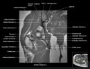
Jonathan mentioned an odd clear gap in the inguinal area, and I wondered if it might partly be due to 'shading' or loss of resolution because of the bulk of muscle and fat in the buttocks.


I'm assuming that's the region I've arrowed here. This is a mid-coronal slice which I've matched approximately to an MR atlas slice. I think the very bright red above and below will be uptake in gluteal and short adductor muscles respectively. The low uptake dark regions will be the femoral heads and greater trochanters, with the femoral necks not fully in-plane.
Jonathan Edwards
Senior Member (Voting Rights)
Jonathan mentioned an odd clear gap in the inguinal area, and I wondered if it might partly be due to 'shading' or loss of resolution because of the bulk of muscle and fat in the buttocks.
I think SNT has covered these things. There are frame artefacts here and calibration issues that may need re-addressing, but taking that on board the pictures still look interesting.
My guess is that this sort of imaging technique can be made generally available if it is really clinically useful. PET scanning has limited applications but some of them are pretty standard now I think. It may still be expensive but prices fall when there is bulk demand.
I get the impression that the images may have been interpreted so far in terms of some clinical concepts that may not be the most relevant - talk of inflammation, mast cells, muscles .... They may be right but clinical input for MECFS studies is often a bit suboptimal!
