Preprint A Novel FiO2 Titration Protocol for Quantifying Pulmonary Oxygen Reserve Capacity : [...], 2025, Qiru

Shiqiru

Established Member
A Novel FiO2 Titration Protocol for Quantifying Pulmonary Oxygen Reserve Capacity : Dynamic Assessment Framework for Infection-Associated Respiratory Dysfunction

Abstract
Background
Progressive decline in pulmonary oxygen reserve capacity (ORC) is a hallmark of infection-associated respiratory dysfunction. Current tools (PaO2/FiO2 ratio, cardiopulmonary exercise testing [CPET], computed tomography [CT]) are limited in dynamic monitoring due to delayed responsiveness, operational complexity, or radiation risks, and other constraints.
Methods The ORC testing methodology integrates the dynamic load-incrementation logic of cardiopulmonary exercise testing (CPET) with the oxygenation quantification framework of PaO2/FiO2. Its operational paradigm comprises three phases:
➀Testing Protocol
Conducted under ventilation-locked conditions, a stepwise FiO2 titration protocol is applied, with termination triggered when SpO2
➁Parameter Definition
The minimum FiO2 required to maintain SpO2 ≥90% (FiO2-MIN) is recorded, and the oxygen reserve capacity is calculated as ORC = 0.21 - FiO2-MIN.
➂Dynamic Modeling
Through continuous monitoring throughout the entire disease course, ORC time-series data are acquired. A time-ORC curve is then fitted, and based on differential calculus (β = ΔORC/Δt, γ = Δβ/Δt), they collectively establish a quantitative respiratory compensation dynamics model in conjunction with the time-ORC curve.
Results The ORC test provides a novel non-invasive tool for dynamic quantification of respiratory reserve. Early warning of ARDS transformation during acute infection and quantitative dynamic tracking of lung dysfunction of long-COVID syndrome are its potential application scenarios. Its clinical utility requires prospective validation through multicenter trials integrated with CPET and CT quantitative analysis.

Link
 

Attachments

Last edited by a moderator:
Hello everyone,
I am the author of the preprint discussing the ORC test. I'd like to take this opportunity to delve deeper into the potential significance of this testing tool.
In my view, the primary bottleneck in current ME/CFS and Long COVID research may not be the choice of research directions (such as viral persistence, autoimmunity, etc.), nor the lack of new microscopic molecular discoveries, or even the absence of novel treatment methods.
The current situation is more akin to astronomy before Kepler – we might not be short of good theories, but we lack the precise, objective observational data needed to validate them, much like the detailed observations and records meticulously collected by Tycho Brahe. Specifically, we are acutely missing quantifiable tools and corresponding continuous data for certain symptoms (which may be driven by specific microscopic molecular mechanisms).
While 2-day CPET is undoubtedly a valuable approach, its limitations and narrow scope of application restrict its broader use. The ORC test, introduced in our preprint, shares conceptual commonalities with CPET (or 6MWT), but it transforms the traditional exercise load challenge into a hypoxic load challenge. We believe this method holds the promise of significantly improving the test's precision, patient tolerability, resistance to interference, and overall applicability.
By continuously plotting the data curve of ORC – an objective, singular indicator – we aim to clearly capture the dynamic changes in the lung's oxygen conversion capacity throughout the disease course. This can provide more precise, objective, and continuous testing data support for future research.
Although preliminary small-scale studies with the ORC test have yielded promising results (I'm happy to share the data if there's interest), I am not a professional researcher in this specific field, and therefore lack the capacity to further expand the test's scope. To fully realize the potential of this testing method, broader collaboration and resources are essential.
Here, I sincerely hope to engage in a comprehensive discussion about this testing method with all of you. My hope is that through collective wisdom, we can collaboratively uncover its utility and explore how to promote its wider application, ultimately contributing to new breakthroughs in the diagnosis and research of ME/CFS and Long COVID.
Thank you again for your attention and valuable insights!
 
It's also worth noting that this preprint is an early version. While the core findings are robust, there are areas where the content and presentation could be improved. Should there be interest, I'd be happy to provide an updated version.
 
While the testing conditions for the ORC method, as described in the preprint, might appear stringent, this is primarily for ensuring methodological standardization and rigor.
In practice, however, much simpler alternative approaches can be adopted for testing. For instance, if one resides in an area with significant altitude variations, such as near mountains, an individual could perform tests at different altitudes using just a fingertip pulse oximeter. This is because changes in altitude directly correspond to changes in oxygen concentration. Thus, altitude itself (or its corresponding oxygen concentration) can directly serve as the testing standard.
Furthermore, the explicit control of ventilation during the described test is also primarily for standardization. In real-world applications, this strict requirement can often be relaxed. Instead, simply performing resting tests after a period of adequate rest each time would suffice.
And should altitude variation not be a feasible option, there are indeed many other alternative methodologies that could be explored. I would be very happy to discuss these possibilities further with anyone interested.
 
Hi and welcome to the forum, @Shiqiru !

The medrxiv formatting is quite difficult to read - I have never understood why they insist on doing it that way. So this might already have been covered in the preprint and I just was unable to find it, but I’m wondering if you could elaborate on which patients you believe this test might be useful for? Long covid is a very broad label that covers anything from critical ICU patients with severe lung damage to someone with just loss of smell 3 months after covid.

PS. Would you be able to add line breaks between the paragraphs in the posts? Many pwME/CFS (including myself) struggle with blocks of text and have to consume written materials in small chunks at a time.
My apologies, I did not include line breaks in my previous response. Thank you very much for the reminder; this is invaluable feedback for me.

A Layman's Explanation of Pulmonary Oxygen Reserve Capacity (ORC):
What is Pulmonary Oxygen Reserve Capacity?


Simply put, it measures your lungs' ability to effectively absorb and deliver oxygen to the bloodstream even when oxygen supply is insufficient (e.g., at high altitudes). It's an indicator of the "redundancy" or "elasticity" of lung function. Just as Cardiopulmonary Exercise Testing (CPET) and the 6-Minute Walk Test (6MWT) assess how the body utilizes this "redundancy" and "elasticity" of the lungs through exercise, the ORC test evaluates it under low-oxygen conditions.

Regarding "Which patients are suitable for ORC testing" and "The broadness of the Long COVID label":

You are absolutely correct that "Long COVID is a very broad label," and not all individuals with Long COVID necessarily experience impaired pulmonary oxygen reserve capacity. However, from our research perspective, as a respiratory infectious disease (and similarly for other respiratory conditions):

Acute Phase: During the acute phase, both severe and mild cases may exhibit a decrease in pulmonary oxygen reserve capacity (ORC), with ORC potentially even becoming negative in severe instances.

Chronic Phase: While symptoms in the chronic phase are diverse, a significant proportion of patients presenting with core symptoms such as fatigue, dyspnea (shortness of breath), and exercise intolerance are highly likely to have abnormalities related to their ORC.

How is the ORC Test Measured, and What are its Units?

The ORC test identifies the critical inspired oxygen concentration at which your blood oxygen saturation (SpO2) begins to fall below 90%.

A lower inspired oxygen concentration at this threshold indicates better pulmonary oxygen reserve capacity.

We quantify this using the formula ORC = 21% - FiO2-MIN, where 21% represents the oxygen concentration in ambient air (21%), and FiO2-MIN is the minimum inspired oxygen fraction when SpO2 drops to 90%. Consequently, the ORC value is expressed as a percentage.

We can draw an analogy with altitude, as higher altitudes naturally entail lower oxygen concentrations:

For a healthy individual at rest, their blood oxygen (SpO2) might remain around 95% (normal) up to approximately 2900 meters (around 9500 feet). If SpO2 begins to drop below 90% at roughly 3000 meters (corresponding to about 15.5% oxygen concentration), their calculated ORC value (via the formula) would be approximately 5.5%.

Conversely, if a patient (e.g., during an acute infection or with pre-existing pulmonary impairment) experiences an SpO2 drop below 90% at an altitude of only 1900 meters (around 6200 feet, corresponding to about 17.2% oxygen concentration), their calculated ORC value would be around 3.8%.

This means that this patient's pulmonary oxygen reserve capacity has decreased by an extent equivalent to 1100 meters of altitude compared to their healthy state. The ORC value has decreased from a healthy 5.5% to 3.8%, a reduction of 1.7 percentage points. This quantitatively indicates an increase in symptom severity, as a lower ORC value signifies more compromised lung function.

Why is ORC Crucial for ME/CFS and Long Covid?

Patients with Myalgic Encephalomyelitis/Chronic Fatigue Syndrome (ME/CFS) frequently suffer from debilitating fatigue, post-exertional malaise (PEM), dyspnea, and exercise intolerance. These pervasive symptoms are intrinsically linked to the body's capacity to acquire and efficiently utilize oxygen. The ORC test offers an objective and quantitative metric that directly reflects the extent of impairment in this crucial physiological function, thereby providing a measurable biological underpinning for these core symptoms.

Pulmonary Oxygen Reserve Capacity (ORC) Testing vs. CPET and 6MWT:

While CPET (Cardiopulmonary Exercise Testing) and 6MWT (6-Minute Walk Test) also provide insights into the body's oxygen utilization capacity, they differ significantly from the ORC test.

CPET and 6MWT evaluate the body's maximal oxygen consumption or exercise endurance by varying physical exertion. They primarily reflect the overall performance of the body's energy systems during an active state.

In contrast, the ORC test assesses this by precisely controlling the inspired oxygen concentration. Its primary focus is on the lungs' adaptive capacity to varying oxygen levels, while also stabilizing pulmonary ventilation changes and eliminating confounding effects typically associated with physical activity.

Advantages of the ORC Test:

Higher Sensitivity: In the 6MWT, many individuals with compromised oxygen reserve capacity may not exhibit a noticeable drop in blood oxygen levels after walking, which can limit the test's diagnostic value. (I believe many 6MWT participants can attest to this experience.) The ORC test, however, can detect subtle reductions in pulmonary oxygen reserve capacity much earlier and with greater precision.

Objective and Quantitative: Any change in ORC is directly reflected and quantified by the ORC test, providing measurable data.
Simpler and More Convenient: Performed in a resting state, the ORC test eliminates the need for strenuous exercise. Its relatively simple equipment also significantly reduces the burden on patients.

Real-world Case Studies:

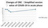
My Wife's Acute COVID-19 ORC Test Curve: On December 6, 2022, my wife tested weakly positive for COVID-19. We initiated simplified ORC testing at 6 PM that day and continued for four consecutive days. By the evening of December 9, although a cough persisted, her other symptoms had significantly improved, so we paused testing. Her antigen test turned negative on December 10. A subsequent test on the evening of December 23 revealed that her pulmonary oxygen reserve capacity had returned to her pre-infection baseline level (as observed on December 6 before the weak positive result).Figure 1

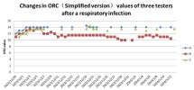
ORC Curves of Three Students After Respiratory Infection: In late November 2023, 4 out of 6 female students in my class's dormitory took leave due to respiratory symptoms, the specific cause of which was not definitively diagnosed. Three of these students, who were proficient in the simplified ORC testing method, verbally agreed to participate in a 5-month ORC tracking study, commencing on November 29.

Based on their ORC curves, their testing likely began after their lowest point of ORC. Students A and C's ORC values recovered to their baseline and stabilized within 4-5 days.

Student B's ORC value, however, after a brief return to baseline, began to fluctuate downward again and consistently remained at a relatively stable, lower level. Despite her subjective feeling of "recovery" and the absence of symptoms other than occasional coughing, her ORC value clearly indicated persistent impairment.Figure 2

This particular case highlights that even when an individual subjectively feels "recovered," objective ORC data can unveil subtle, underlying manifestations of post-infection sequelae. Student B's ORC curve serves as a potential quantifiable indicator for changes in post-infectious symptoms. (Notably, Student B's ORC eventually returned to the baseline levels of Students A and C by February 13, 2024, though this data is not depicted in the current figures, it can be provided.)

Potential Value of ORC Testing for ME/CFS and Long COVID Research:

Based on the cases presented, I trust that ORC testing is now more clearly understood. As a singular, objective, and precise indicator of pulmonary oxygen reserve capacity, it offers distinct advantages. For patients with conditions like Long COVID or ME/CFS who experience related symptoms such as fatigue, dyspnea, and exercise intolerance, ORC testing offers:

Condition Monitoring: Regular ORC testing (e.g., every other day or at longer intervals) can provide insight into the progression of their condition, even in the absence of a pre-illness baseline. An increasing ORC value suggests improvement, while a decreasing value may indicate worsening.

Treatment Efficacy Evaluation: Current evaluations of treatment outcomes often rely on subjective patient reports, which lack precision. The ORC test can provide a precise, objective, and quantifiable standard for assessing treatment effectiveness, aiding in the determination of the most beneficial therapeutic approaches.

As I've emphasized, converting subjective, ambiguous assessments into precise, objective quantitative metrics is an indispensable prerequisite for advancements in this field. While the ORC test may only address the symptom spectrum related to pulmonary dysfunction, it has the potential to be a transformative 'game-changer,' serving as an objective benchmark for various aspects of ME/CFS and Long COVID research.
 

Attachments

  • 2.jpg
    2.jpg
    49.3 KB · Views: 4
  • 3.jpg
    3.jpg
    66.6 KB · Views: 4
@Jaybee00 This is also a long process, as can be seen from my response, the relevant research has gone through a long time. Fortunately, after actively searching, I came here. I hope my findings and research can be useful.
 
Thank you for the post and additional detail. Reading the preprint, I understood that this may be relevant in two scenarios. The first is to try and determine people at risk for ARDS/SIRS (adult respiratory distress syndrome / systemic inflammatory response syndrome) in the acute setting. Potentially allowing something like baricitinib or steroids to arrest the descent into needing invasive mechanical ventilation. As you write, someone on the verge of intensive care isn't going to be doing a CPET so an alternative would be helpful. Often once the more severe pathophysiology is recognised it's been a rapid change, but too late and you're committed to a rough ICU course of life-support; where your technique could give earlier warning.

If I've understood correctly, in simple terms this looks like evaluating the effect of increased inspired oxygen levels when someone is on supplementary O2 and inverting the paradigm, testing what happens when it's lower than normal atmosphere (21%). I imagine healthy people wouldn't be particularly bothered down to say 16% for example over a testing period. But someone who was borderline in terms of physiologic reserve might observably drop their sats even at a modest reduction to 19-20% FiO2.

The second is the chronic scenario, where lung function is deteriorating but not yet clinically overt. I think this would tend to be things like the developing pulmonary fibrosis side of long COVID. The question for us here is whether it relates to the more ME/CFS type of LC. There have been some studies showing pulmonary impairment in the absence of CT changes like ground glass opacities and fibrosis.


Maybe if it's a feature only of LC though I think I recall seeing some mild drop-offs in SpO2 during six-minute walk tests in some studies. We do also have some indicators pointing to RBCs and haemoglobin in ME/CFS, though personally I wondered if the problem might more relate to oxygen delivery and usage (eg at muscles) rather than uptake in lungs. (In my first year of LC, but no longer permanently bedbound I was recording extremely low SvO2 of 22% and even 16% while visiting ICU colleagues on their blood gas analyser, despite perfectly normal SpO2). So I think it's a good idea to try and understand what's happening with oxygen utilisation in ME/LC beyond respiratory oxygen input, ie are mitochondria using too much O2 (inefficiently)?

I appreciate you want to keep your test non-invasive, using medical-grade pulse oximeters, rather than needing a blood draw, but have you considered incorporating peripheral SvO2 in your evaluation? An alternative non-invasive peripheral measure that could be part of your non-invasive test might be muscle NIRS. See my historical comments on SvO2 and SmO2.
Thank you for your detailed and insightful comments! I'm very pleased that you've grasped the nuances of our proposed ORC framework so well. Your interpretation of the first scenario (acute early warning) is spot on. As you rightly pointed out, CPET is unsuitable for critically ill patients, and our method aims to provide an earlier, safer, and more feasible assessment tool. This could potentially buy crucial time for clinical decisions (such as when to initiate interventions like steroids or baricitinib), thereby possibly preventing the need for more invasive treatments.

Regarding the second scenario (chronic lung dysfunction), your understanding is again excellent. While pulmonary fibrosis is indeed a potential cause for abnormal ORC, we believe that more pervasive mechanisms in these conditions often involve microvascular dysfunction, microclot formation, and potentially reduced red blood cell deformability. These microscopic pathophysiological changes, even in the absence of overt findings on routine CT scans, can lead to a sustained decrease in pulmonary gas exchange efficiency, manifesting as persistently sub-baseline ORC. This is highly relevant to the common symptoms of post-exertional malaise (PEM) and fatigue experienced by individuals with Long COVID and ME/CFS.

Your mention of experiencing extremely low SvO2 (22% and even 16%) with perfectly normal SpO2 during your first year of Long COVID is indeed a highly concerning observation, and it directly reflects the issues with oxygen delivery and utilization that you suspected. I am very troubled to hear this and truly hope your condition has improved. Your experience perfectly highlights the critical distinction between our proposed ORC test and SvO2:

  • The ORC test is specifically designed to purely assess the lung's intrinsic ability to transfer oxygen from inhaled air into the bloodstream (i.e., its gas exchange efficiency and reserve). It focuses on the 'entry point' of oxygen into the body – the pulmonary level of oxygenation.

  • In your case, with perfectly normal SpO2, even without any additional physiological load (such as external oxygen consumption from exercise), your SvO2 was extremely low. This suggests that while your lungs' ability to pick up oxygen at rest was adequate, the issue lies further downstream – in the systemic circulation, specifically regarding oxygen delivery to and utilization by the tissues. This could involve insufficient cardiac output, microcirculatory dysfunction (systemic, not exclusively pulmonary), mitochondrial dysfunction leading to inefficient oxygen utilization, or excessive oxygen extraction by tissues.
Therefore, incorporating peripheral SvO2 (e.g., via femoral vein) or muscle NIRS into the assessment is indeed highly valuable for patients like yourself, who present with normal SpO2 but underlying systemic issues with oxygen delivery/utilization. These measures directly reflect tissue oxygenation and oxygen extraction rates.

As you astutely pointed out, post-exertional malaise and fatigue are often the ultimate manifestation of complex interactions within the human body, with oxygen uptake (conversion/transfer) and consumption (utilization) at their core. Given that most Long COVID/ME/CFS patients maintain normal resting SpO2, this strongly suggests that the underlying oxygenation issues frequently emerge under physiological load, or indicate an inadequate oxygen reserve capacity.

This is precisely where the innovation and groundbreaking nature of our ORC test lies. By simulating a controlled hypoxic load, the ORC test directly quantifies the lung's capacity to effectively convert and transfer oxygen into the bloodstream – its true pulmonary oxygen reserve. With this tool, we can more precisely diagnose and differentiate the root causes of exercise intolerance and fatigue: Is it a 'supply' problem due to insufficient pulmonary oxygen transfer (uptake) capacity, or a 'demand' problem stemming from inefficient systemic oxygen consumption (utilization) or excessive extraction by tissues, or a combination of both?

Based on our research principles and existing preliminary observations, we hypothesize that abnormalities in pulmonary oxygen transfer (i.e., reduced ORC) may be far more prevalent than currently recognized, simply because they are often not detectable by routine clinical assessments that lack a controlled load component. This also implies that the incidence of symptomatic oxygen transfer abnormalities might be more widespread than commonly assumed.

Therefore, while complementary measures like CPET and muscle NIRS are undeniably valuable for assessing systemic oxygen delivery and utilization, the ORC test offers a unique and indispensable perspective, allowing us to more precisely identify the 'bottleneck' within the oxygen metabolic chain—specifically, the limitations in pulmonary oxygenation reserve.

We envision that the ORC test could, in the future, serve as an integral component within CPET or other comprehensive physiological assessment protocols, or be conducted separately alongside measures like muscle NIRS for mutual corroboration. Through such a multi-modal, integrated assessment, we aim to comprehensively and fully characterize the pathophysiological landscape of Long COVID/ME/CFS patients, thereby providing more targeted foundations for diagnosis and subsequent therapeutic interventions.

Looking ahead, once the ORC test gains widespread adoption, its distinct advantages as a singular, objective, precise, and longitudinally trackable indicator of pulmonary oxygen transfer reserve will become increasingly apparent. It is poised to become a fundamental cornerstone for related research, empowering deeper exploration into disease mechanisms and more rigorous evaluation of intervention efficacy.
 
My apologies, I did not include line breaks in my previous response. Thank you very much for the reminder; this is invaluable feedback for me.
Thank you for being so considerate and for the thorough explanation!
Chronic Phase: While symptoms in the chronic phase are diverse, a significant proportion of patients presenting with core symptoms such as fatigue, dyspnea (shortness of breath), and exercise intolerance are highly likely to have abnormalities related to their ORC.
Why is ORC Crucial for ME/CFS and Long Covid?

Patients with Myalgic Encephalomyelitis/Chronic Fatigue Syndrome (ME/CFS) frequently suffer from debilitating fatigue, post-exertional malaise (PEM), dyspnea, and exercise intolerance. These pervasive symptoms are intrinsically linked to the body's capacity to acquire and efficiently utilize oxygen.
Are these claims substantiated by the evidence?

I’m intrigued by the possibility of measurements that can be used even when bedridden without causing harm. But I also know that my experience with dyspnea, exercise intolerance and PEM isn’t really like how I felt at the summit of Mt. Kilimanjaro, where I presumably had quite low SpO2, or when skiing at high altitudes in the Alps.

Fatigue is a very general symptom that certainly can be cause by peripheral issues, but it can presumably also be caused without any of those issues as well. After all, it only exists in the brain.
 
Thanks @Shiqiru , I am familiar with the physiology but tend not to keep abbreviations in my head.

My main concern is that we don't have good evidence for symptoms in MECFS being related to oxygen availability or other aspects of "energy metabolism". There is no clinical evidence of poor oxygenation of tissues as far as I know. CPET has been talked about a lot but not because it is abnormal per se in MECFS but that it shows a change on a second test on a second day. Thus may have some relation to symptoms of PEM but that is far from clear and it would not be a simple explanatory relation.

In general people with MECFS do not complain of dyspnoea on exertion. The symptoms that get called fatigue do not seem likely to be due to hypoxia per se and are not the same as the various normal usages of "fatigue". Even those are generally independent of dyspnoea and hypoxia.

There has been a lot of stuff on Xitter about microclots but there is no clinical sign of any microthrombi and the lab data are pretty uninterprerable and unconvincing. In short, I don't see any good reason to link MECFS to tissue hypoxia at present and suspect that the method is only relevant to acute lung damage as in acute Covid, or subsequent scarring.
 
Thank you for being so considerate and for the thorough explanation!


Are these claims substantiated by the evidence?

I’m intrigued by the possibility of measurements that can be used even when bedridden without causing harm. But I also know that my experience with dyspnea, exercise intolerance and PEM isn’t really like how I felt at the summit of Mt. Kilimanjaro, where I presumably had quite low SpO2, or when skiing at high altitudes in the Alps.

Fatigue is a very general symptom that certainly can be cause by peripheral issues, but it can presumably also be caused without any of those issues as well. After all, it only exists in the brain.
Thank you for leaving another message. That experience on Mt. Kilimanjaro and skiing in the Alps sounds amazing. It's truly regrettable that your current illness affects you so profoundly.

Regarding "Are these claims substantiated by the evidence?"

To answer this question, we need to look at it from a few perspectives:

1. Our Preliminary Test Results:

Although we are using a simplified version that may have a larger margin of error, the preliminary results are very encouraging:

The ORC values of healthy test subjects are almost a flat line, indicating that it is a very stable physiological indicator for a healthy baseline.
During the acute phase of respiratory infections (whether worsening or recovering), the ORC curves show extreme smoothness and consistency, sensitively reflecting the degree of symptom changes.

For patients with potential chronic conditions, during stable periods, the ORC curve also approaches a straight line.

These findings suggest that ORC is a very stable indicator for healthy individuals. Furthermore, for respiratory infections or chronic lung dysfunction, it is a very sensitive and reliable indicator capable of reflecting the extent of symptom changes. This gives ORC tremendous potential to become a reliable core indicator in relevant research.

2. Theoretical Basis:

Theoretically, ORC is derived from the application of the oxygenation index (PaO₂/FiO₂) and its combination with CPET. Scholars in related fields can easily understand its principles. Of course, from a rigorous perspective, ORC testing still requires further validation. At present, we urgently need more people to understand it, so that further validation and research work can be carried out.

Regarding High-Altitude Hypoxia-Induced Sensations and Infection-Related Fatigue:

Concerning the sensations caused by high-altitude hypoxia and the issue of post-infection fatigue, I would like to say:

While fatigue certainly has a subjective component, it is a fundamental fact that high altitudes (low oxygen) lead to a more severe sense of fatigue after exercise compared to lower altitudes. Even without exercise, higher altitudes can cause fatigue. These phenomena are all related to changes in oxygen conversion efficiency.

However, I personally cannot fully empathize with the precise feeling you describe when comparing this sensation to PEM.

The Relationship Between ORC, Fatigue, and PEM:
It's important to clarify:

An abnormal ORC does not necessarily mean there will be a feeling of fatigue similar to PEM. This was evident in the case of student B: her ORC remained abnormal, but she did not subjectively report significant fatigue.

PEM does not necessarily mean ORC will be abnormal. From an oxygen perspective, PEM results from the combined effect of both oxygen supply (input) and oxygen utilization (output).

In other words, ORC is not a direct measure of fatigue, but it is closely related to fatigue (especially exercise intolerance and post-exertional fatigue).

The Great Potential of ORC Testing and Practical Advice:

From my perspective, a significant proportion of ME/CFS and Long COVID patients may have ORC values somewhat below the baseline for their age group (ORC naturally decreases with age).

ORC is a relatively easy-to-obtain test data. As I mentioned before, if you are in an area with mountains or plateaus, you can try self-testing with a pulse oximeter.

If, while resting, your blood oxygen saturation starts to drop below 90% at altitudes below 2500 meters (for individuals under 50 years old), it likely means your ORC is below the normal baseline.

If you have the means and are interested, I highly encourage you to try it.

If this can be substantiated in a significant proportion of patients, then ORC has the potential to become a core biomarker for quantitative research. Its unique strength lies in being a single, objective, precise, and easily measurable indicator. While the range of symptoms it directly relates to might be relatively narrow (primarily linked to lung function and oxygen utilization), it could be a key to unlocking the underlying pathological mechanisms.

For an individual, seeing their ORC gradually recover through treatment and rehabilitation would also be very encouraging.
 
Thanks @Shiqiru , I am familiar with the physiology but tend not to keep abbreviations in my head.

My main concern is that we don't have good evidence for symptoms in MECFS being related to oxygen availability or other aspects of "energy metabolism". There is no clinical evidence of poor oxygenation of tissues as far as I know. CPET has been talked about a lot but not because it is abnormal per se in MECFS but that it shows a change on a second test on a second day. Thus may have some relation to symptoms of PEM but that is far from clear and it would not be a simple explanatory relation.

In general people with MECFS do not complain of dyspnoea on exertion. The symptoms that get called fatigue do not seem likely to be due to hypoxia per se and are not the same as the various normal usages of "fatigue". Even those are generally independent of dyspnoea and hypoxia.

There has been a lot of stuff on Xitter about microclots but there is no clinical sign of any microthrombi and the lab data are pretty uninterprerable and unconvincing. In short, I don't see any good reason to link MECFS to tissue hypoxia at present and suspect that the method is only relevant to acute lung damage as in acute Covid, or subsequent scarring.
Thank you for your candid feedback and for clarifying your familiarity with the physiology while preferring fewer abbreviations. I will try to be mindful of that.

You raise a crucial point about the perceived lack of direct evidence linking ME/CFS symptoms to oxygen availability or other aspects of "energy metabolism." I believe this perception often stems from the fact that common measures like SpO2 (either at rest or during a 6-Minute Walk Test with an insufficient load) typically appear normal. This can lead to a misunderstanding that oxygenation is unaffected, when in fact, the compensatory oxygen reserve capacity (ORC) might already be compromised.

Regarding CPET and Evidence for Oxygen/Energy Metabolism Links:

Regarding CPET, the meta-analysis "Use of Cardiopulmonary Exercise Testing to Evaluate Long COVID-19 Symptoms in Adults" indeed shows a significant reduction in peak V̇O2 among Long COVID patients, alongside "peripheral limitations (abnormal oxygen extraction)." This indicates that the combined process of oxygen acquisition and consumption is impaired. As I've discussed, the issue could be with acquisition, consumption, or both. ORC, as a single indicator focusing on the lung's reserve capacity to manage oxygen under stress, could help clarify this. Incorporating ORC testing into such studies would illuminate the specific contribution of this reserve capacity.

ORC as a Monitoring Tool:

Furthermore, the progression of ME/CFS and Long COVID symptoms often isn't sudden. ORC could be an excellent tool for monitoring these subtle changes. As illustrated by student B's case, she was unaware of her abnormal ORC. While her symptoms hadn't overtly worsened, ORC indicated a physiological deficit. If symptoms were to further deteriorate, it's likely that not only ORC but other physiological aspects would also be compromised, at which point symptoms would become noticeable.

ORC and 2-Day CPET:

Indeed, the 2-day CPET, which reveals post-exertional malaise (PEM) after high-intensity exercise, is widely considered the most reliable method for assessing ME/CFS and Long COVID. I hypothesize that if ORC testing were integrated into a 2-day CPET protocol, we would very likely observe a significant decline in ORC on the second day as well, serving as another objective marker of PEM.

Dyspnea and ORC:

You're right that individuals with ME/CFS may not typically complain of dyspnea on exertion. However, this does not necessarily mean their ORC is undamaged. I can only reiterate that if feasible (e.g., if you live near mountains or high-altitude areas), trying a simple self-test with a pulse oximeter could be insightful.

Microclots and Measurement Tools:

Regarding microclots and impaired red blood cell deformability – while there might not be easily identifiable corresponding symptoms at present, I believe this is precisely because we lack appropriate measurement tools to bridge the micro and macro levels. Testing ORC could potentially establish a crucial link between cellular-level damage (like microclots) and observable, albeit subtle, physiological symptoms captured by ORC changes.
 
Your responses look increasingly like AI products @Shiqiru and that puzzles me. To be honest I don't see any of this adding up in relation to MECFS. Fatigue in MECFS is not the fatigue of normal parlance. Poor oxygenation is one of a hundred causes of symptoms called fatigue but until there is more convincing evidence I see no reason to thi k it has anything to do with MECFS.

For a theory to work you need to consider all the things that don't fit as well as the ones that might fit. To me the things that don't fit loom much larger when it comes to implicating energy metabolism, and especially oxygen access in MECFS.

As an anecdote, I regularly ski at 3500 meters in the Alps and walk at up to 5000 metres in the Andes. I don't recognize any particular relation between altitude and fatigue, just breathlessness and "the wall".
 
My main concern is that we don't have good evidence for symptoms in MECFS being related to oxygen availability or other aspects of "energy metabolism". There is no clinical evidence of poor oxygenation of tissues as far as I know. CPET has been talked about a lot but not because it is abnormal per se in MECFS but that it shows a change on a second test on a second day. Thus may have some relation to symptoms of PEM but that is far from clear and it would not be a simple explanatory relation.

In general people with MECFS do not complain of dyspnoea on exertion. The symptoms that get called fatigue do not seem likely to be due to hypoxia per se and are not the same as the various normal usages of "fatigue". Even those are generally independent of dyspnoea and hypoxia.

This is not in-line with my experience. I had a one off CPET on two separate occasions. The second one being at Papworth Hospital.

My VO2 was 19.7 ml/kg/min (68% predicted for my age and weight) at maximum and 11.2 ml/kg/min at my anaerobic threshold.

Several years before that my VO2 max was 45% of predicted despite my RER being over 1 demonstrating I put in enough effort. I don’t believe this is a result of pure deconditioning.

I also got a poor diastolic and systolic blood pressure response during exercise despite being on Midodrine. I’ve been diagnosed with SFN and “POTS” too which I know you feel doesn’t mean much.

I get incredibly breathless on exertion. Worse when my ME is more severe. I pushed myself to my limit to get to the CPETs to demonstrate disability despite being wheelchair bound.

I’m of the understanding that Dr Systrom’s invasive CPET data proves problems on day 1 too not just day 2. I understand we need suitable controls etc but I don’t think this kind of physiological disturbance can be overlooked.

Does the above mean to say I don’t have ME yet I fulfil every current diagnostic criteria or do but with an extra co-morbidity or I’m of a specific subset? I know this can’t be answered but I know I’m not alone in my experience either.
 
@Shiqiru I think there might be a misunderstanding about the nature of ME/CFS and PEM.

While PEM literally means post-exertional malaise, it’s so much more than just feeling less well. It’s a distinct pattern of increased symptoms, new symptoms and the worsening of functioning that is often delayed by 12-48 hours after what would otherwise be trivial exertion or stimuli.

This period can last for days, weeks or sometimes months, and is not alleviated by rest or sleep.

To put it in layman’s terms: it feels like waking up being hit by a truck, having a terrible flu, the worst hangover in your life and having ran a marathon the day before. After taking a shower sitting down the day before.

It’s completely disproportional to the trigger(s), and most pwME/CFS describe it as unlike anything they’ve ever experienced before, including other severe illnesses and post-viral fatigue.

I have no doubt that oxygen issues can cause fatigue and other symptoms, but I struggle to see how that has anything to do with ME/CFS.

We have recently produced a factsheet on PEM if you’re interested in more details:
 
Back
Top Bottom