Differentiating ME/CFS Through Raman Spectroscopic Profiling of Immune Cells
Anuradha Ramoji, Frida Naumann, Tze Ching Yeo, Sabine Vonderlind, Aikaterini Pistiki, Hulya Yilmaz, Iwan W. Schie, Oleg Ryabchkov, Thomas Bocklitz, Andreas Stallmach, Jürgen Popp, Philipp Reuken
[Line breaks added]
Background
ME/CFS is a systemic, debilitating disease of unknown cause and pathophysiology. It is characterized by physical and mental fatigue with wide range of clinical manifestations. A hallmark symptom of ME/CFS is post-exertional malaise (PEM) and individuals with COVID-19 history reported similar clinical features, suggesting common underlying mechanisms between ME/CFS and post-viral syndromes.
Current diagnostic criteria rely heavily on clinical judgement, leading to potential delays and misdiagnosis. To address this gap, our study uses Raman spectroscopy, a label-free optical technique capable of detecting molecular changes in cells, as a potential diagnostic platform for ME/CFS with PEM.
Specifically, we focus on PBMCs to detect molecular changes associated with ME/CFS with PEM before and after exercise, we aim to determine whether Raman spectroscopic signatures can reliably discriminate immune cell status associated with symptom exacerbation.
Methods
Cohort: 18 ME/CFS patients with PEM and 16 healthy control
Samples: 9 ml EDTA blood collected before (visit 1) and 24h after (visit 2) m1’SST physical exertion., Immune cells from blood: PBMCs
Method: Raman spectroscopy with 785nm excitation laser, ~ 2000 cells/visit, 1s/spectrum/cell, 60x WI objective
Results

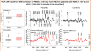
➢ The PCA-LDA coefficients suggest biochemical changes in immune cells allow their differentiation from visit-1 to visit-2.
➢ The Raman model was able differentiate with accuracy ~70% the PBMCs from patient, whereas the model cannot differentiate PBMCs from healthy controls before and after physical exertion
➢ The prediction accuracy increased to ~80% for PBMCs when seperate models where built for the different subgroups of patients (grouped based on the difference spectrum shown in the right figure)
Conclusion
The present study illustrates the role of the immune system in the pathophysiology of ME/CFS with PEM. The results of a predictive model based on Raman data indicate immunological changes in patients with long COVID.
The ability to discriminate between patients before and after physical exertion demonstrates the influence of minimal physical activity on their immune system. Thus, Raman spectroscopy is a promising diagnostic platform and can aid in the diagnosis of ME/CFS with PEM.
Furthermore, two subgroups were identified, reflecting the high clinical heterogeneity of the disease. The identification and study of the different subgroups could provide crucial information about different biochemical changes of cells and consequently different manifestations and severity of symptoms.
As a result, Raman spectroscopy could open up new opportunities for research into the aetiology and pathophysiology of CFS and long-term COVID.
Poster (Some more charts on the poster)
Anuradha Ramoji, Frida Naumann, Tze Ching Yeo, Sabine Vonderlind, Aikaterini Pistiki, Hulya Yilmaz, Iwan W. Schie, Oleg Ryabchkov, Thomas Bocklitz, Andreas Stallmach, Jürgen Popp, Philipp Reuken
[Line breaks added]
Background
ME/CFS is a systemic, debilitating disease of unknown cause and pathophysiology. It is characterized by physical and mental fatigue with wide range of clinical manifestations. A hallmark symptom of ME/CFS is post-exertional malaise (PEM) and individuals with COVID-19 history reported similar clinical features, suggesting common underlying mechanisms between ME/CFS and post-viral syndromes.
Current diagnostic criteria rely heavily on clinical judgement, leading to potential delays and misdiagnosis. To address this gap, our study uses Raman spectroscopy, a label-free optical technique capable of detecting molecular changes in cells, as a potential diagnostic platform for ME/CFS with PEM.
Specifically, we focus on PBMCs to detect molecular changes associated with ME/CFS with PEM before and after exercise, we aim to determine whether Raman spectroscopic signatures can reliably discriminate immune cell status associated with symptom exacerbation.
Methods
Cohort: 18 ME/CFS patients with PEM and 16 healthy control
Samples: 9 ml EDTA blood collected before (visit 1) and 24h after (visit 2) m1’SST physical exertion., Immune cells from blood: PBMCs
Method: Raman spectroscopy with 785nm excitation laser, ~ 2000 cells/visit, 1s/spectrum/cell, 60x WI objective
Results

➢ The PCA-LDA coefficients suggest biochemical changes in immune cells allow their differentiation from visit-1 to visit-2.
➢ The Raman model was able differentiate with accuracy ~70% the PBMCs from patient, whereas the model cannot differentiate PBMCs from healthy controls before and after physical exertion
➢ The prediction accuracy increased to ~80% for PBMCs when seperate models where built for the different subgroups of patients (grouped based on the difference spectrum shown in the right figure)
Conclusion
The present study illustrates the role of the immune system in the pathophysiology of ME/CFS with PEM. The results of a predictive model based on Raman data indicate immunological changes in patients with long COVID.
The ability to discriminate between patients before and after physical exertion demonstrates the influence of minimal physical activity on their immune system. Thus, Raman spectroscopy is a promising diagnostic platform and can aid in the diagnosis of ME/CFS with PEM.
Furthermore, two subgroups were identified, reflecting the high clinical heterogeneity of the disease. The identification and study of the different subgroups could provide crucial information about different biochemical changes of cells and consequently different manifestations and severity of symptoms.
As a result, Raman spectroscopy could open up new opportunities for research into the aetiology and pathophysiology of CFS and long-term COVID.
Poster (Some more charts on the poster)
