Myopathy as a cause of Long COVID fatigue: Evidence from quantitative and single fiber EMG and muscle histopathology, April 2023, Hejbøl et al

EndME

Senior Member (Voting Rights)
Myopathy as a cause of Long COVID fatigue: Evidence from quantitative and single fiber EMG and muscle histopathology

Highlights
Objective
To describe neurophysiological abnormalities in Long COVID and correlate quantitative electromyography (qEMG) and single fiber EMG (sfEMG) results to clinical scores and histopathology.

Methods
84 patients with non-improving musculoskeletal Long COVID symptoms were examined with qEMG and sfEMG. Muscle biopsies were taken in a subgroup.

Results
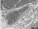
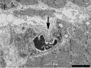
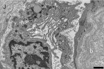
Mean motor unit potential (MUP) duration was decreased in ≥ 1 muscles in 52 % of the patients. Mean jitter was increased in 17 % of the patients in tibialis anterior and 25 % in extensor digitorum communis. Increased jitter was seen with or without myopathic qEMG. Low quality of life score correlated with higher jitter values but not with qEMG measures. In addition to our previously published mitochondrial changes, inflammation, and capillary injury, we show now in muscle biopsies damage of terminal nerves and motor endplate with abundant basal lamina material. At the endplate, axons were present but no vesicle containing terminals. The post-synaptic cleft in areas appeared atrophic with short clefts and coarse crests.

Conclusions
Myopathic changes are common in Long COVID. sfEMG abnormality is less common but may correlate with clinical scores. sfEMG changes may be due to motor endplate pathology.


https://www.sciencedirect.com/science/article/pii/S1388245723000196#b0040

This seems to build on similar work discussed here: https://www.s4me.info/threads/myopa...uscle-histopathology-2022-hejbøl-et-al.27997/.
 
Interested to see if and how this might change in the long term.

From their 2022 paper

10 biopsies presented signs of mitochondrial involvement as COX-negative fibers, abnormal mitochondrial structure, or larger subsarcolemmal mitochondria accumulations

Which sounds a lot like Mitochondrial abnormalities in the postviral fatigue syndrome (1991, Acta Neuropathologica) —

Groups of mitochondria, together with the nucleus and abundant glycogen, were present in a sub-sarcolemmal position and also scattered between muscle fibres with their long axis parallel to that of the fibres. They showed severe pleomorphism of shape and size

The other point made in their 2022 paper was —

Empty capillary basal membranes were found in three cases, indicating capillary loss

We keep seeing this.

Most remarkable were the two patients who after 10–13 months showed extensive basal lamina production, associated not only with muscle fibers but also with capillaries and nerves. We additionally observed mitochondrial pathology and capillary changes, the latter suggesting ongoing capillary remodeling processes. The importance of the vascular mechanisms in COVID-19 pathology has been described in previous studies. One possible reason for the capillary degeneration could be the presence of occluding thrombocyte aggregations. Microthrombi have previously been found in acute COVID-19. In this context, our finding of capillary segments with unusual accumulation of Weibel–Palade bodies, known to be essential for platelet adhesion, is interesting. Another factor could be remodeling induced by fiber atrophy.

Rob Wüst presented at the recent Charité conference on LC and said

"And super provocatively, the other thing I would like to discuss is this image you see on the right. These are microclots inside skeletal muscle - and they are not inside capillaries."

In the picture he showed they looked to be where the capillaries would previously have been, so maybe they were in empty capillary basal membranes?
 
This is also what I'd be interested in. Can these dust looking like microclots and similar problems still occur in these muscle biopsies if one is looking inside of skeletal muscles?

I guess it'd be a lot clearer if they weren't present in samples from a control group processed at the same time. Haven't the energy to read the article properly, so I'm not sure whether they did this.
 
I guess it'd be a lot clearer if they weren't present in samples from a control group processed at the same time. Haven't the energy to read the article properly, so I'm not sure whether they did this.

I don't think that'll be possible since Rob Wüst's findings haven't been published (the article is about a different study). It's more of a question for those that have knowledge of such biopsies without being able to read the study.
 
Last edited:
We previously found myopathic changes in quantitative electromyography (qEMG) in 11 of 20 patients referred for electrophysiological investigation with suspicion of polyneuropathy due to paresthesia (Agergaard et al., 2021) and suggested that myopathy might explain physical fatigue. Recently we published light and electron microscopy results of muscle biopsies from 16 Long COVID patients with fatigue, in which we found pathological findings in all patients including microvascular changes as well as affection of the myofibrils with mitochondrial changes, atrophy, inflammation and multiple basal membrane as a sign of regeneration (Hejbøl et al., 2022).

In order to further explore whether myopathy might be a common finding in Long COVID patients, we aimed to examine a larger cohort of Long COVID patients in a Post COVID Clinic with fatigue and musculoskeletal symptoms (Agergaard et al., 2022). We aimed also to examine a possible neuromuscular transmission failure which could be a cause of fatigue. We here present the qEMG and single fiber EMG (sfEMG) investigations of patients referred for electrophysiological evaluation from a Post COVID Clinic and describe their clinical and electrophysiological characteristics including presentation of histology in a small subgroup of these patients.


Forum threads on these three previous papers by the same Danish team:
Myopathic changes in patients with long-term fatigue after COVID-19, 2021, Agergaard et al

Myopathy as a cause of fatigue in long-term post-COVID-19 symptoms: Evidence of skeletal muscle histopathology, 2022, Hejbøl et al

Characteristics of a Danish Post COVID Cohort referred for examination due to persistent symptoms 6 months after... COVID-19, 2022, Agergaard et al
 
Last edited:
qEMG - quantitative electromyography
A review of clinical quantitative electromyography, 2010
"Information regarding the morphology of motor unit potentials (MUPs) and motor unit firing patterns can be used to help diagnose, treat, and manage neuromuscular disorders. In a conventional electromyographic (EMG) examination, a clinician manually assesses the characteristics of needle-detected EMG signals across a number of distinct needle positions and forms an overall impression of the condition of the muscle. Such a subjective assessment is highly dependent on the skills and level of experience of the clinician, and is prone to a high error rate and operator bias.

Quantitative methods have been developed to characterize MUP waveforms using statistical and probabilistic techniques that allow for greater objectivity and reproducibility in supporting the diagnostic process."


sfEMG - single fibre electromyography
Single-fiber EMG: A review, 2011
"When a motor axon is depolarized the action potentials travel distally and excite the muscle fiber more or less at the same time. The variation in the time interval between the two action potentials of the same motor unit is called as “jitter”. SFEMG measures the variation of this inter potential interval (jitter)[Figure 3]."

Screen Shot 2023-07-22 at 2.05.13 pm.png
 
Is it right to say that it can't be due to the processing method of the samples? Which is often a criticism of microclots in the blood.

One of the criticisms about ex vivo microclot demonstration was that blood likes to make clots, particularly at room temperature. Although we only had 12 seconds of video comment, the point was made that these were amyloid deposits, not in blood vessels, therefore not in blood. The image on screen showed these as green fluorescent deposits, so possibly using the amyloid tagging Thioflavin-T that Resia Pretorius has published on. I think it's been said that that can stick to quite a lot of other stuff, so there is still room for it being artifact.

Rob Wüst characterised them as "microclots", but not blocking capillary flow. The question that I hope will be addressed in their upcoming paper is whether they think they were previously in capillaries and so previously obstructed flow — and required the capillary remodelling discussed in this thread's papers. I have no idea whether it would be possible to separate them off and compare their proteomics to the Pretorius findings.

It's more of a question for those that have knowledge of such biopsies without being able to read the study.

I'll try and ask a pathology colleague about this, though we may need to wait for the paper.
 
From December 2020 to March 2022 a total of 659 patients were examined in the Post COVID clinic and 107 patients were received for EMG investigation[*]. In all patients, qEMG and sfEMG were performed, and 99 patients accepted the use of their clinical data for this research study. From the 99 patients, 15 were excluded due to: other diseases with risk of or proven peripheral neuropathy (diabetes in three patients, neuropathy in five patients and hereditary neuropathy in one patient), no positive SARS-CoV-2 test (two patients), qEMG investigation less than 12 weeks after initial COVID-19 (one patient), age below 18 (one patient), or insufficient investigation due to patient compliance (two patients). Thus, 84 patients had qEMG performed and were included in analyses.

*referred for electromyography (EMG) examination due to severely affected and non-improving daily function, as well as physical/muscular exhaustion, myalgia or reduced force.

76% of patients female
16% have abnormal levels of creatine kinase (indicates muscle damage)
8% have abnormal levels of myoglobulin (indicates muscle damage)
For both creatine kinase and myoglobulin, the abnormal values were only slightly above the normal reference value.

95% have physical fatigue
88% have myalgia
92% have concentration difficulties
55% have reduced muscle force

Investigations around 10 months after acute Covid-19
None of the 84 patients had mononeuropathy or entrapment neuropathy in peroneal nerve or signs of polyneuropathy. None of the patients had decrement in repetitive nerve stimulation.

I think these videos are useful for understanding this paper; they cover motor neurons and the motor endplate (in the muscle around the terminal of the motor neuron) and action potentials.

 
Last edited:
Discussion said:
This study showed myopathic changes in qEMG and/or increased jitter in sfEMG in 53 of the 84 patients (63 %) referred to electrophysiological testing for neuromuscular Long COVID symptoms. Myopathic qEMG was more common (52 %) than sfEMG (23 %), and seven of the 84 patients (8 %) showed both. There was no correlation between the clinical scores and qEMG measures while more pronounced sfEMG changes correlated with some of the more severe Long COVID symptom scores. Muscle biopsy in a subgroup showed as previously published (Hejbøl et al., 2022) a wide variety of histological findings including mitochondrial changes, inflammation, and capillary injury. The novelty in the present study is that we show now damage of terminal nerves and motor endplate.


Discussion said:
The myopathic changes in the patients with only abnormal sfEMG suggest that the primary pathology may be in the muscle fibers. In the present study, up to 76 % of the patients had increased polyphasia on qEMG, which may be explained by unequal muscle fiber diameter due to muscle fiber atrophy or changes of regeneration such as basal lamina duplications detected by histology. We also showed in our previous study uncommon amounts of basal lamina not only surrounding muscle fibers but also around nerves and capillaries.

Basal lamina?
google said:
The basement membrane, also known as basal lamina, is a thin, dense layer of extracellular matrix that lines most human tissues forming the supporting structure and scaffolding for epithelial tissue and separates different types of cells, such as nerve cells and muscle cells.

Discussion said:
In one patient with myopathic qEMG but normal sfEMG, we were lucky to visualise the motor endplate in electron microscopy with abundant basal lamina material and atrophic post synaptic cleft. These changes may suggest a similar mechanism in patients with normal as well as abnormal sfEMG. Most interestingly, the sfEMG measures correlated with the severity of clinical scores as MUP durations did not. From these results, one may speculate that abnormalities in motor endplate might be giving most of the symptoms, and the patients particularly with abnormal sfEMG may benefit from medications such as Pyridostigmine.

Pyridostigmine? (Mestinon)
google said:
Pyridostigmine has been used therapeutically to improve muscle strength in patients with myasthenia gravis, a chronic autoimmune neuromuscular disease characterized by skeletal muscle weakness. Pyridostigmine, used intravenously, may reverse the effects of neuromuscular blocking agents.

The authors acknowledge the small number of biopsies.
 
I guess it'd be a lot clearer if they weren't present in samples from a control group processed at the same time. Haven't the energy to read the article properly, so I'm not sure whether they did this.
The EMG stuff was compared against previously derived values for people of the same age and sex. So, I think when they say a result is abnormal, they have some basis for saying that.

The histology is a bit beyond me. They are saying that in the images of nerve terminals that they captured from one biopsy, the "boutons", the swellings right at the end, are missing. I've tried looking at the paper in the picture and at pictures of normal motor nerve terminals and, yeah, ... maybe? It would be good to hear what people who routinely look at these sort of images make of them.
 
Back
Top Bottom